Magnetic Resonance Imaging

Magnetic resonance imaging (MRI) is the gold standard imaging modality for soft tissues. It produces detail cross-sectional images of soft tissue and bone anatomy, including muscles, tendons, ligaments, brain and organs, without the use of ionizing radiation. In addition to orthopedic imaging, MRI is also used for heart, brain and breast. MRI uses gadolinium contrast in many exams to highlight tissues and blood vessels, which enhances images and offers better diagnostic quality. It can also be used in conjunction with PET scans. How does MRI work? MR creates images by using powerful magnets to polarize hydrogen atoms in water (the body is made of of more than 80% water) so they face in one direction. A radiofrequency pulse is then used to ping these atoms, causing them to wobble, or resonate. The MRI coils detect this and computers can assemble images from the signals. Basic MRI scans will focus on the resonance of fat and water in two different sequences, which highlight and contrast different features in the anatomy.

Experts develop cardiac MRI reference values for healthy adolescents

The newly developed reference values could have “direct implications for clinical practice” and can be used by any provider to determine whether an adolescent falls within the normal range of measurements for their age group.

Providers still routinely using unnecessary imaging to diagnose mild traumatic brain injury

There is little value in deploying CT, MRI or X-ray to diagnose mTBI, experts note, and it may instead result in harm and excess costs.

Thumbnail

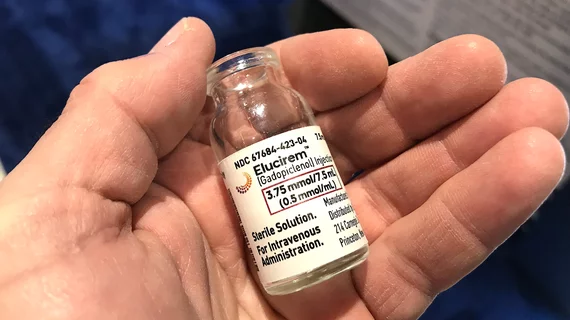
GE HealthCare's new MRI contrast agent advances to clinical trial

Experts are hopeful that the first-of-its-kind injectable manganese-based macrocyclic agent will offer a suitable alternative to GBCAs, as it can be more efficiently eliminated from the body.

Thumbnail

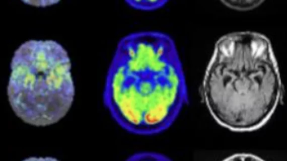
MRI findings differ significantly in boys and girls who have sustained concussions

Findings from functional MRI brain scans following concussions in pediatric patients indicate that injury recovery times may differ between sexes. 

Thumbnail

Rod Stewart is helping to address 'ridiculous' backlogs by paying for patients' imaging exams

The legendary singer's actions were prompted, in part, by his own experience undergoing medical imaging recently.

Thumbnail

Insights into early MRI-termination risk factors offer helpful strategies for prevention

Even a small number of early terminations can be costly, in addition to creating negative experiences for patients, experts wrote in the European Journal of Radiology. 

Thumbnail

MRI catches CT in head-to-head lung imaging

When it comes to assessing patients with suspected pulmonary embolism, contrast-enhanced CT pulmonary angiography has no diagnostic edge over a certain free-breathing, unenhanced MRI perfusion protocol.

0.55T MRI lumbar compared to 1.5T sequence #lumbarMRI #1.5T

Low-field MRI systems improve accessibility, but what kind of diagnostic quality do they offer?

Experts recently compared the use of a 0.55T low-field MRI system to a 1.5T system to determine whether the results rendered were within an acceptable diagnostic range.