Magnetic Resonance Imaging

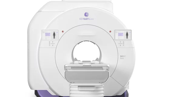
Magnetic resonance imaging (MRI) is the gold standard imaging modality for soft tissues. It produces detail cross-sectional images of soft tissue and bone anatomy, including muscles, tendons, ligaments, brain and organs, without the use of ionizing radiation. In addition to orthopedic imaging, MRI is also used for heart, brain and breast. MRI uses gadolinium contrast in many exams to highlight tissues and blood vessels, which enhances images and offers better diagnostic quality. It can also be used in conjunction with PET scans. How does MRI work? MR creates images by using powerful magnets to polarize hydrogen atoms in water (the body is made of of more than 80% water) so they face in one direction. A radiofrequency pulse is then used to ping these atoms, causing them to wobble, or resonate. The MRI coils detect this and computers can assemble images from the signals. Basic MRI scans will focus on the resonance of fat and water in two different sequences, which highlight and contrast different features in the anatomy.

MRI exams reveal the impact of steroid use on the brain

Experts involved in the study suggest that their findings could help explain the neuropsychiatric effects seen in many patients who are prescribed steroids long-term.

WTC first responders

Can PTSD cause dementia? Imaging of 9/11 first responders suggests it's possible

Compared to World Trade Center responders who have not been diagnosed with PTSD, those who had received the diagnosis were observed to have decreased fractional anisotropy of white matter tracts on their brain MRIs.

Thumbnail

5 imaging modalities have changed the war on cancer over the past 50 years. How many can you name?

Two branch chiefs with the cancer imaging program at NIH’s National Cancer Institute have produced a written commemoration to mark the golden anniversary of 1971’s National Cancer Act.

Poor midlife health leads to advanced brain aging, MRI analysis shows

Though the participants’ actual ages ranged from 69 to 72 years old, the experts’ predictive analysis revealed their estimated current brain ages to be between 46 and 93.

idiopathic intracranial hypertension

MRI a useful tool for avoiding invasive procedures for suspected idiopathic intracranial hypertension

The experts maintained that these MRI parameters support the diagnosis of IIH in clinically suspicious cases and could help reduce the use of invasive procedures when aligned with symptoms.

Thumbnail

Machine learning model uses MRI data to identify candidates for liver transplant

When applied to MRI and clinical features, different machine learning models were recently shown to reliably predict post-treatment recurrences of hepatocellular carcinoma. 

Thumbnail

Deep learning system outperforms rads in brain tumor identification and classification

The new findings findings represent “a step toward improved tumor diagnoses," according to authors of a new study published in JAMA.