Magnetic Resonance Imaging

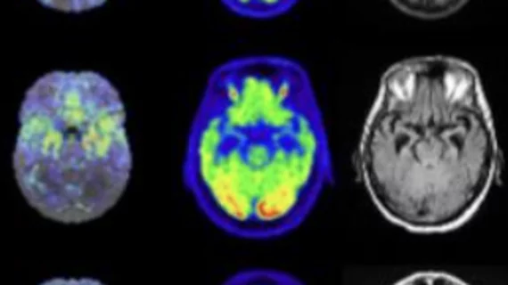
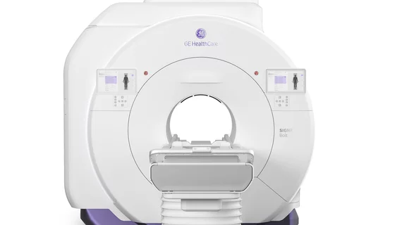
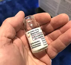
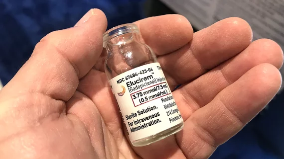
Magnetic resonance imaging (MRI) is the gold standard imaging modality for soft tissues. It produces detail cross-sectional images of soft tissue and bone anatomy, including muscles, tendons, ligaments, brain and organs, without the use of ionizing radiation. In addition to orthopedic imaging, MRI is also used for heart, brain and breast. MRI uses gadolinium contrast in many exams to highlight tissues and blood vessels, which enhances images and offers better diagnostic quality. It can also be used in conjunction with PET scans. How does MRI work? MR creates images by using powerful magnets to polarize hydrogen atoms in water (the body is made of of more than 80% water) so they face in one direction. A radiofrequency pulse is then used to ping these atoms, causing them to wobble, or resonate. The MRI coils detect this and computers can assemble images from the signals. Basic MRI scans will focus on the resonance of fat and water in two different sequences, which highlight and contrast different features in the anatomy.

CMR of a patient showing evidence of myocardial hypertrophy

Cardiac MRI findings linked with worse outcomes in hypertension patients

Nonischemic LGE was revealed to be independently associated with worse outcomes in hypertension patients. 

Radiology, meet ‘3D-based superconducting radiofrequency computers’

What do you get when you combine MRI with quantum computing? The world will soon find out.

Technologist ‘learning opportunities’ vastly outnumber imaging ‘do-overs’ across almost 1 million exams

Reviewing a 20-month run with a radiologist-to-technologist communications tool, researchers have found minor problems with image quality 10 times more common than patient callbacks for repeat imaging.

New MRI findings suggest that even moderate drinkers are at risk of alcohol-related cognitive decline

In the largest known study to date on the relationship between moderate alcohol consumption and iron homeostasis, experts suggested that as little as four drinks per week can increase the presence of iron deposits on imaging.

Thumbnail

Imaging-based COVID study finds blood clots twice as likely with infection vs. vaccination

A retrospective analysis of cross-sectional venograms shows only 4.5% of a teaching hospital’s COVID-vaccinated patients had a clotting condition in early 2021. Meanwhile the rate for the unvaccinated was 10.1%.

Startup OEM expands pediatric use cases for portable MRI

Portable MRI maker Hyperfine is touting a new capability in its Swoop scanner that may appeal to pediatric neurologists and neurosurgeons as well as neuroradiologists.

Thumbnail

New MRI method can identify early signs of Parkinson's

The researchers are now working on developing a qMRI method that can be incorporated into clinical settings, with an anticipated timeline of three to five years. 

Imaging surveillance program detects pancreatic cancer in its earliest stages

Individuals at high risk of pancreatic cancer benefit from annual imaging and have decreased mortality rates compared to those who forgo preventive screening.