Magnetic Resonance Imaging

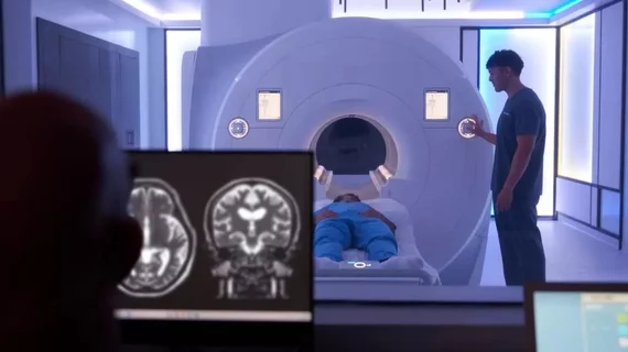
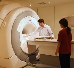
Magnetic resonance imaging (MRI) is the gold standard imaging modality for soft tissues. It produces detail cross-sectional images of soft tissue and bone anatomy, including muscles, tendons, ligaments, brain and organs, without the use of ionizing radiation. In addition to orthopedic imaging, MRI is also used for heart, brain and breast. MRI uses gadolinium contrast in many exams to highlight tissues and blood vessels, which enhances images and offers better diagnostic quality. It can also be used in conjunction with PET scans. How does MRI work? MR creates images by using powerful magnets to polarize hydrogen atoms in water (the body is made of of more than 80% water) so they face in one direction. A radiofrequency pulse is then used to ping these atoms, causing them to wobble, or resonate. The MRI coils detect this and computers can assemble images from the signals. Basic MRI scans will focus on the resonance of fat and water in two different sequences, which highlight and contrast different features in the anatomy.

Political ideologies have distinct connectivity signatures on MRI, research shows

Can a person’s political affiliation be identified based on imaging of their brain, without them ever saying a word or starting a debate on social media? The answer might surprise many. 

Cardiac MRI findings can predict incident CVD years before onset

Prior imaging of the descending thoracic and abdominal aorta in study participants revealed thoracic aortic wall area (AWA), plaque prevalence and plaque volumes to be independently associated with incident CVD.

Neighbor to the North facing a ‘very bleak future’ if medical imaging not modernized soon

Canada has fallen behind other industrialized nations in multiple measures of capital healthcare investment, and the most glaring gap is found in the state of its medical imaging equipment.

Thumbnail

New MRI technique a ‘virtual biopsy’ for surveilling transplanted hearts

Researchers have developed a novel cardiovascular MRI protocol as an option to the invasive gold standard, endomyocardial biopsy, for monitoring heart-transplant patients at risk of suffering organ rejection.

Photo and caption courtesy of American Journal of Roentgenology (AJR), American Roentgen Ray Society (ARRS). Used with permission.

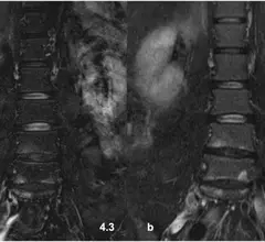
Diffusion-weighted MRI boosts detection of locally recurrent pancreatic cancer

MRI with DWI images showed higher sensitivity in detecting local recurrence after PDAC resection.

Doctor patient with masks

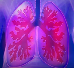
New imaging technique detects post-COVID lung abnormalities

Conventional imaging did not detect the abnormalities, which MRI scans identified up to one year after infection.

Some respiratory face masks are unsafe for MRIs, study finds

Five out of eight filtering face piece (FFP-3) masks studied contained ferromagnetic components.

MRI contact-sport study shows ‘no concussion’ doesn’t mean ‘no brain changes’

Football players whose heads are repeatedly struck but suffer no concussions have white-matter abnormalities similar to those sustained by their concussed peers.