Magnetic Resonance Imaging

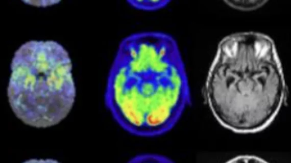
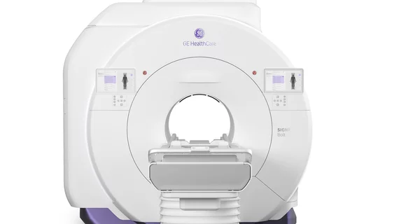
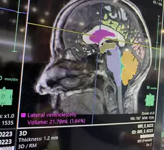
Magnetic resonance imaging (MRI) is the gold standard imaging modality for soft tissues. It produces detail cross-sectional images of soft tissue and bone anatomy, including muscles, tendons, ligaments, brain and organs, without the use of ionizing radiation. In addition to orthopedic imaging, MRI is also used for heart, brain and breast. MRI uses gadolinium contrast in many exams to highlight tissues and blood vessels, which enhances images and offers better diagnostic quality. It can also be used in conjunction with PET scans. How does MRI work? MR creates images by using powerful magnets to polarize hydrogen atoms in water (the body is made of of more than 80% water) so they face in one direction. A radiofrequency pulse is then used to ping these atoms, causing them to wobble, or resonate. The MRI coils detect this and computers can assemble images from the signals. Basic MRI scans will focus on the resonance of fat and water in two different sequences, which highlight and contrast different features in the anatomy.


Thumbnail

Top MRI trends include helium-free systems, compressed sensing and AI

Signify Research outlines several key technologies and trends in MRI to look for at the Radiological Society of North America 2022 meeting.

Philips running short on MRI coil disposables, FDA advising providers to consider image-guidance alternatives

In a letter to providers, the agency recommends Philips clients in the affected product category consider any of three steps to cope with the interruption. 

Thumbnail

How maternal COVID-related stress can alter fetal brain development

The preliminary results of a new study published in the medRxiv preprint server highlight variances in fetal brain development based on mothers’ perceived stress during the onset of COVID.

X-rays may suffice for assessing some low-back fractures

It’s unlikely radiography will unseat MRI for routinely distinguishing between acute and chronic compression fractures of the lumbar vertebrae, but certain X-ray findings might, in cases, obviate the need for the pricier imaging option.

Thumbnail

The brain imaging-based argument for letting kids play video games

Functional MRI brain imaging studies have revealed higher brain activity associated with attention and memory for gamers who play three or more hours per day. 

MRI shows structural, functional brain abnormalities in Lyme disease long-haulers

Compared to a group of patients who had not been previously diagnosed with Lyme disease, those who had been infected displayed unusual activity in the frontal lobe of the brain on functional MRI scans.

Structural brain abnormalities linked to functional seizures

Using MRI imaging, the experts found that patients who experienced functional seizures had thinning in the superior temporal cortex and thickening of the left occipital cortex.

FDA greenlights AI-powered MR software that could give radiotherapy planning a boost

The U.S. Food and Drug Administration granted Philips 510(k) clearance for its AI-powered MRI platform tailored to the treatment of head and neck cancers.