Magnetic Resonance Imaging

Magnetic resonance imaging (MRI) is the gold standard imaging modality for soft tissues. It produces detail cross-sectional images of soft tissue and bone anatomy, including muscles, tendons, ligaments, brain and organs, without the use of ionizing radiation. In addition to orthopedic imaging, MRI is also used for heart, brain and breast. MRI uses gadolinium contrast in many exams to highlight tissues and blood vessels, which enhances images and offers better diagnostic quality. It can also be used in conjunction with PET scans. How does MRI work? MR creates images by using powerful magnets to polarize hydrogen atoms in water (the body is made of of more than 80% water) so they face in one direction. A radiofrequency pulse is then used to ping these atoms, causing them to wobble, or resonate. The MRI coils detect this and computers can assemble images from the signals. Basic MRI scans will focus on the resonance of fat and water in two different sequences, which highlight and contrast different features in the anatomy.

Thumbnail

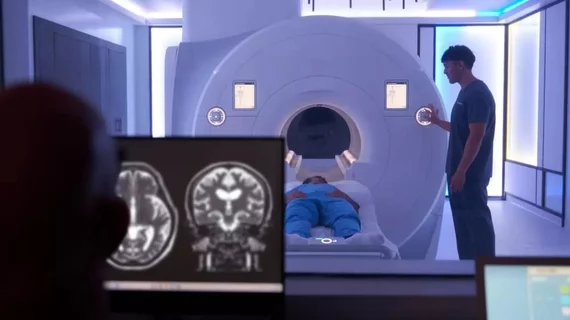
A ‘magical’ experience: Patients given psychedelics don’t mind MRI noise, constraints

A new clinical trial in Australia is one of the largest in the world to use brain imaging to test the impact of psychedelics on neural activity.

brain scans of woman who died after taking experimental alzheimer's drug

Brain scans of patient who died while on experimental Alzheimer's drug cause experts to question safety

A neurologist who examined the patient's imaging explained that her brain swelling was so severe in some cases that the folds of the cerebral cortex appeared “merged and squashed.”

Cross-organ imaging illuminates the heart-brain-liver axis

A population-level study featuring multi-organ MRI has confirmed that problems in any of three major organs—the heart, brain or liver—tend to co-occur with unfavorable findings in either or both of the other two.

Thumbnail

Radiologist- and technologist-related errors to blame for most musculoskeletal MRI recalls

These errors are frequently due to a breakdown in communication between radiologists, technologists and referring providers, either before or during exams, according to a new analysis of more nearly 63,000 musculoskeletal MRI exams.

Sean Fain, PhD, vice chair of radiology and research and a professor of radiology, Division of Cardiovascular and Thoracic Imaging, University of Iowa, discusses how long-COVID lung damage can be tracked using xenon (Xe) gas magnetic resonance imaging (MRI) and quantitative computed tomography (CT). He spoke to Health Imaging at RSNA 2022.

VIDEO: Tracking long-COVID lung damage using MRI and CT

Sean Fain, PhD, vice chair of radiology and research and a professor of radiology, University of Iowa, discusses how long-COVID lung damage can be tracked using xenon (Xe) gas MRI and quantitative CT at RSNA 2022. 

Imaging industry names in the news: Koning, Medality, QT Imaging, Rezolut, Scanslated, more

Noteworthy market developments listed in the order announcements were posted.    

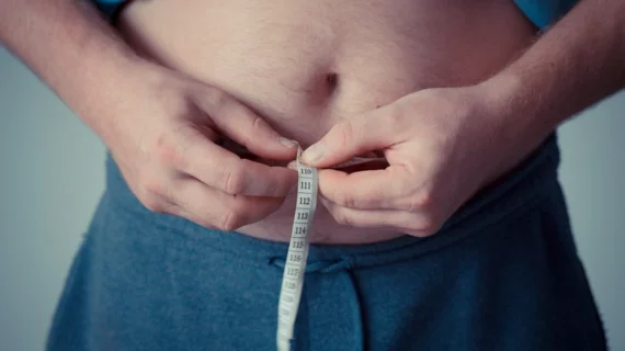
Why are women at lower risk for brain inflammation? Subcutaneous fat may hold the key

Researchers examined male and female mice on high-fat diets to learn about the impacts of fat distribution on brain inflammation. 

prostate cancer PSA

On its own, MRI-targeted biopsy takes big bite out of prostate cancer overdiagnosis

The gain came with the delayed discovery of only a few clinically significant cancers.