Magnetic Resonance Imaging

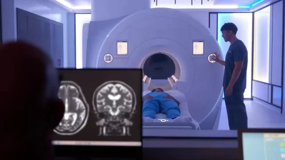
Magnetic resonance imaging (MRI) is the gold standard imaging modality for soft tissues. It produces detail cross-sectional images of soft tissue and bone anatomy, including muscles, tendons, ligaments, brain and organs, without the use of ionizing radiation. In addition to orthopedic imaging, MRI is also used for heart, brain and breast. MRI uses gadolinium contrast in many exams to highlight tissues and blood vessels, which enhances images and offers better diagnostic quality. It can also be used in conjunction with PET scans. How does MRI work? MR creates images by using powerful magnets to polarize hydrogen atoms in water (the body is made of of more than 80% water) so they face in one direction. A radiofrequency pulse is then used to ping these atoms, causing them to wobble, or resonate. The MRI coils detect this and computers can assemble images from the signals. Basic MRI scans will focus on the resonance of fat and water in two different sequences, which highlight and contrast different features in the anatomy.

dementia alzheimer's brain

MRI scans suggest anti-amyloid drugs can cause accelerated brain volume loss

In some cases, certain classes of anti-amyloid beta (Aβ) drugs resulted in individuals’ brain volume decreasing more rapidly while using the drug than they would have if they had not received treatment at all. 

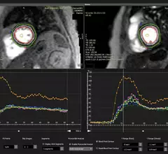
Medtronic just launched its MRI Care Pathway, a new system that can streamline the process of completing MRI scans for patients with Medtronic MRI compatible cardiac devices.

Medtronic streamlines MRI screening, scanning for patients with implanted cardiac devices

Medtronic launched its MRI Care Pathway, a new system that can streamline the process of completing MRI scans for patients with compatible cardiac devices.

Female Medical Research Scientist Working with Brain Scans

Neuroradiologists make more mistakes when facing these 3 scenarios

“These findings should be considered when designing workflow-related and other interventions seeking to reduce errors," experts wrote in AJR

quality

MRI protocol tweak nets big savings for hospital system while opening up additional exam slots

An inordinate number of patients with metallic hardware were receiving inappropriate MRIs without a key sequence to reduce related artifacts. 

Thumbnail

Radiologists outperform commercially available AI in PI-RADS scoring

The findings contradict prior research that utilized the same software, experts involved in the research noted. This could be due to out-of-distribution data for the DL software, which could impair its performance. 

Thumbnail

Cindy Crawford-backed, whole-body MRI startup continues expansion, despite radiologist concern

California-based Prenuvo recently raised $70 million and is opening several locations across the U.S. this year and next. 

Details emerge on yet another MRI accident

The accident happened on February 23, and it has since been revealed that it occurred at Kaiser Permanente’s Redwood City Medical Center facility.

Iodine contrast being loaded into a contrast injector in preparation for a cardiac CT scan at Duly Health and Care in Lisle, Illinois. The contrast shortage is causing some healthcare organizations to postpone exams and procedures and ration contrast supplies. Photo by Dave Fornell

How to manage patients in need of contrast-enhanced imaging following an allergic-like reaction

Up to one-third or more of patients who experience moderate or severe allergic-like reactions to ICM must have their imaging protocols revised when subsequent exams involving contrast are requested.