Magnetic Resonance Imaging

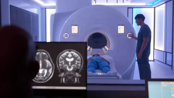
Magnetic resonance imaging (MRI) is the gold standard imaging modality for soft tissues. It produces detail cross-sectional images of soft tissue and bone anatomy, including muscles, tendons, ligaments, brain and organs, without the use of ionizing radiation. In addition to orthopedic imaging, MRI is also used for heart, brain and breast. MRI uses gadolinium contrast in many exams to highlight tissues and blood vessels, which enhances images and offers better diagnostic quality. It can also be used in conjunction with PET scans. How does MRI work? MR creates images by using powerful magnets to polarize hydrogen atoms in water (the body is made of of more than 80% water) so they face in one direction. A radiofrequency pulse is then used to ping these atoms, causing them to wobble, or resonate. The MRI coils detect this and computers can assemble images from the signals. Basic MRI scans will focus on the resonance of fat and water in two different sequences, which highlight and contrast different features in the anatomy.

 background parenchyma enhancement breast MRI

MRI finding linked to heightened cancer risk among women with very dense breasts

A new AI model helped researchers identify a relationship between background parenchyma enhancement on breast MRI exams and breast cancer risk.

Use of advanced imaging skyrockets in ED, and radiologists should be ready for more

The surge in imaging utilization is independent of increased patient volumes, “suggesting that other extrinsic drivers” contributed, researchers noted. 

EHR nudge helps health system drop use of unnecessary lumbar X-rays by 53%

Researchers with NYC Health + Hospitals detailed their large-scale quality improvement project in the Journal of the American College of Radiology.

Demand for imaging to swell after approval of new Alzheimer’s drug, GE HealthCare predicts

"We believe that this is a pretty profound growth opportunity across the space," CEO Peter Arduini told investors during a Q2 earnings call. 

liver cancer

Incorrect CT protocol leads to radiologist overlooking liver cancer, patient’s untimely death

The physician had assigned the standard, four-phase protocol for liver imaging, but the radiographer instead performed a single, portal-venous phase only.

Thumbnail

PROMISE trial results reinforce confidence in new MRI contrast agent

The randomized, double-blinded phase 3 study revealed gadopiclenol to be noninferior to gadobutrol for all qualitative visualization parameters as rated by all blinded readers involved in the trial.

Thumbnail

Experts argue for the use of MRI in all first-episode psychosis patients

A new meta-analysis revealed that up to 26% of FEP patients display intracranial radiological abnormalities on MRI, some of which can alter clinical management.

Money bankruptcy debt liquidation

Imaging vendor ViewRay files for Chapter 11 bankruptcy

The Denver-based company is letting go more than 100 staffers, including its CEO, interim CFO and chief legal officer.