Magnetic Resonance Imaging

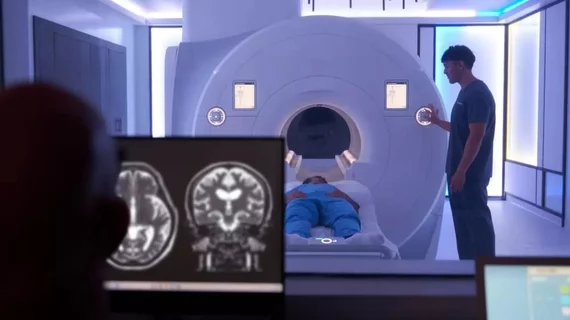
Magnetic resonance imaging (MRI) is the gold standard imaging modality for soft tissues. It produces detail cross-sectional images of soft tissue and bone anatomy, including muscles, tendons, ligaments, brain and organs, without the use of ionizing radiation. In addition to orthopedic imaging, MRI is also used for heart, brain and breast. MRI uses gadolinium contrast in many exams to highlight tissues and blood vessels, which enhances images and offers better diagnostic quality. It can also be used in conjunction with PET scans. How does MRI work? MR creates images by using powerful magnets to polarize hydrogen atoms in water (the body is made of of more than 80% water) so they face in one direction. A radiofrequency pulse is then used to ping these atoms, causing them to wobble, or resonate. The MRI coils detect this and computers can assemble images from the signals. Basic MRI scans will focus on the resonance of fat and water in two different sequences, which highlight and contrast different features in the anatomy.

MRA for pulmonary embolus

MR angiography a suitable alternative to CT when ruling out pulmonary embolus

The modality switch became especially important during the iodinated contrast shortage of 2022 when clinics were forced to deploy mitigation tactics as a means of preserving their contrast supply. 

Thumbnail

Radiologists read across an average of 5 subspecialties but aren’t always confident doing so

About 40% of rads said they accept studies across all areas of expertise. Yet, less than half are “very confident” reading them. 

FDA greenlights software that supports blood-flow analysis via MRI without contrast

iTFlow enables the visualization of fluid dynamics in the heart and blood vessels in both 2D and 3D, developer Cardio Flow Design said Monday.

SimonMed

Radiology practice SimonMed launches nationwide whole-body MRI service, charging fraction of competitors’ prices

SimonOne utilizes AI, advanced clinical protocols and the “best available” equipment to detect diseases before symptoms arise, the PE-backed group announced. 

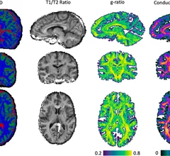
New MRI technique offers detailed view of COVID's impact on the brain

The new technique mixes MRI signals at different gradient pulse strengths and timings, offering better visualization of differences in the way water molecules move through tissues.  

Thumbnail

When GBCA use in musculoskeletal imaging is and is not appropriate

Although gadolinium-based contrast agents are largely considered safe and are routinely used for MRI exams, experts suggest that providers should still utilize GBCAs sparingly for musculoskeletal studies.

Thumbnail

Prostate cancer size is frequently underestimated on imaging

Newly published data recently revealed just how common the extent of prostate cancer is underestimated based on imaging measurements. 

University of Minnesota team develops compact portable MRI

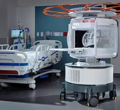
Curbside MRI: Experts are developing system small enough to fit in a pickup

Someday, getting an MRI exam could be as simple as having food delivered to your door—at least that is the hope of a group of experts at the University of Minnesota who are working on a compact system said to be small enough to sit in the bed of a truck.