Magnetic Resonance Imaging

Magnetic resonance imaging (MRI) is the gold standard imaging modality for soft tissues. It produces detail cross-sectional images of soft tissue and bone anatomy, including muscles, tendons, ligaments, brain and organs, without the use of ionizing radiation. In addition to orthopedic imaging, MRI is also used for heart, brain and breast. MRI uses gadolinium contrast in many exams to highlight tissues and blood vessels, which enhances images and offers better diagnostic quality. It can also be used in conjunction with PET scans. How does MRI work? MR creates images by using powerful magnets to polarize hydrogen atoms in water (the body is made of of more than 80% water) so they face in one direction. A radiofrequency pulse is then used to ping these atoms, causing them to wobble, or resonate. The MRI coils detect this and computers can assemble images from the signals. Basic MRI scans will focus on the resonance of fat and water in two different sequences, which highlight and contrast different features in the anatomy.

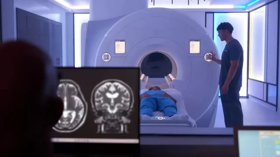
The European Society of radiology European Congress of Radiology (ECR) 2023 meeting. Image courtesy of ECR

Key trends in radiology at the European Congress of Radiology 2023 meeting

Bhvita Jani, research manager at the healthcare market analysis firm Signify Research, shares noteworthy happenings from the ECR expo floor.

Thumbnail

Experts identify sex-specific MRI brain signatures associated with obesity

Researchers at the University of California Los Angeles combined multimodal brain MRIs with clinical and personal data from 183 men and women to identify sex-specific brain signatures that could be indicative of what causes a person to develop obesity.

portable MRI in pediatric ECMO setting

Portable MRI achieves 'immense success' in study of critically ill children

Results from the world’s first study on the use of portable MRI equipment during pediatric ECMO therapy were published April 4 in Critical Care.

PET/MRI of FAPI radiotracer uptake in Crohn's disease

New imaging technique could change how Crohn's disease is treated

The FAPI radiotracer experts used in this research can bind specifically to the connective tissue cells that cause intestinal wall fibrosis common to Crohn's disease.

vaccine syringe covid-19 coronavirus

Shoulder injury related to vaccine administration: How the latest MRI data describe the condition

 Although rare, SIRVA made its way into headlines following the widespread rollout of COVID vaccines. This prompted a renewed push among the medical community to better define the side effect.

dementia alzheimer's brain

MRI scans suggest anti-amyloid drugs can cause accelerated brain volume loss

In some cases, certain classes of anti-amyloid beta (Aβ) drugs resulted in individuals’ brain volume decreasing more rapidly while using the drug than they would have if they had not received treatment at all. 

Medtronic just launched its MRI Care Pathway, a new system that can streamline the process of completing MRI scans for patients with Medtronic MRI compatible cardiac devices.

Medtronic streamlines MRI screening, scanning for patients with implanted cardiac devices

Medtronic launched its MRI Care Pathway, a new system that can streamline the process of completing MRI scans for patients with compatible cardiac devices.

Female Medical Research Scientist Working with Brain Scans

Neuroradiologists make more mistakes when facing these 3 scenarios

“These findings should be considered when designing workflow-related and other interventions seeking to reduce errors," experts wrote in AJR