Magnetic Resonance Imaging

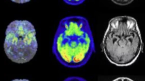
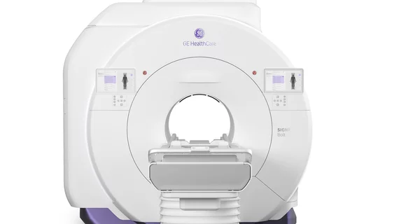
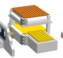
Magnetic resonance imaging (MRI) is the gold standard imaging modality for soft tissues. It produces detail cross-sectional images of soft tissue and bone anatomy, including muscles, tendons, ligaments, brain and organs, without the use of ionizing radiation. In addition to orthopedic imaging, MRI is also used for heart, brain and breast. MRI uses gadolinium contrast in many exams to highlight tissues and blood vessels, which enhances images and offers better diagnostic quality. It can also be used in conjunction with PET scans. How does MRI work? MR creates images by using powerful magnets to polarize hydrogen atoms in water (the body is made of of more than 80% water) so they face in one direction. A radiofrequency pulse is then used to ping these atoms, causing them to wobble, or resonate. The MRI coils detect this and computers can assemble images from the signals. Basic MRI scans will focus on the resonance of fat and water in two different sequences, which highlight and contrast different features in the anatomy.

Thumbnail

Breast MRIs are on the rise—but not among women who need them most

Communities are failing to follow guidelines that ensure women at an increased risk for breast cancer receive additional MRI screening, according to research from the University of New Mexico’s School of Medicine.

Thumbnail

Civic official says ‘overburdened staff’ at least partly responsible for MRI-related death in India

A 32-year-old man died on Jan. 27 after being sucked into an MRI chamber at BYL Nair Hospital in Mumbai, India. The civic investigation into the man’s death has not yet released its official report, but a senior official with knowledge of the investigation said at least part of the blame goes to “overburdened staff and facilities at hospitals.”

Thumbnail

New cardiac MRI process can improve results, ease restrictions on patients

A team of researchers from Cedars-Sinai Medical Center in Los Angeles have developed a new method for cardiac MRI that can be complete in just 90 seconds. The technique also allows patients to breathe during testing, with promises of more accurate, more reliable results.

Thumbnail

How a Michigan practice cut its MRI wait times from 101 days to 5

As radiology practices grapple with the specialist shortages and drawn-out wait times that accompany a nationwide push for more specialized MRI, one Michigan practice is successfully solving both problems.

Thumbnail

Offshoot of digital mammography could provide alternative to breast MRI

An adaptation of digital mammography could cut image acquisition times and provide a new screening method to patients unable to undergo breast MRI, but added time commitments could make the technique difficult to implement in clinical practice, a case study published in the Journal of the American College of Radiology suggests.

Thumbnail

Increased background parenchymal enhancement on screening MRI associated with higher risk of breast cancer

High-risk patients with any amount of background parenchymal enhancement (BPE) beyond the minimum on screening MRI have an increased risk of developing breast cancer, according to a new study published by Academic Radiology.

Thumbnail

Spanish team applies MR imaging to quality control in the meat industry

Scientists at the University of Extremadura in Badajoz, Spain, have found another use for MR imaging—but it has nothing to do with medicine, the Spanish Foundation for Science and Technology has announced.

Thumbnail

UK officials worry patients are getting 'too big' for standard MRI machines

In one of Western Europe’s most overweight areas, British officials are struggling to tackle a growing obesity epidemic that’s barring hundreds of patients from receiving MRI exams, the BBC reported this week.