Magnetic Resonance Imaging

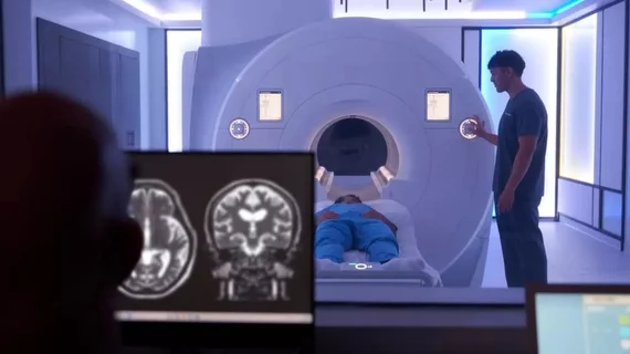
Magnetic resonance imaging (MRI) is the gold standard imaging modality for soft tissues. It produces detail cross-sectional images of soft tissue and bone anatomy, including muscles, tendons, ligaments, brain and organs, without the use of ionizing radiation. In addition to orthopedic imaging, MRI is also used for heart, brain and breast. MRI uses gadolinium contrast in many exams to highlight tissues and blood vessels, which enhances images and offers better diagnostic quality. It can also be used in conjunction with PET scans. How does MRI work? MR creates images by using powerful magnets to polarize hydrogen atoms in water (the body is made of of more than 80% water) so they face in one direction. A radiofrequency pulse is then used to ping these atoms, causing them to wobble, or resonate. The MRI coils detect this and computers can assemble images from the signals. Basic MRI scans will focus on the resonance of fat and water in two different sequences, which highlight and contrast different features in the anatomy.

Thumbnail

Boston hospital installs first MRI system of its kind, says new 7T scanner now available

Brigham and Women’s Hospital (BWH) in Boston made two big imaging-related announcements this week. First, it has installed the first NICU-dedicated MRI system in the U.S. Second, its 7T scanner is now officially available for patients.

Thumbnail

Inner peace: MRI scans suggest mindful people may feel less pain

MRI scans show that more mindful individuals may feel less pain.

Thumbnail

‘Father of MRI’ receives posthumous award for contributions to health imaging

Paul Lauterbur, MD, a former chemistry professor at Stony Brook University in New York known as the “Father of MRI,” was posthumously awarded the Long Island Section of the Institute of Electrical and Electronics Engineers (IEEE) Historical Milestone Plaque for his contributions to the development of MRI.

Thumbnail

Which breast MRI findings can predict lymphovascular invasion in cancer patients?

Tumor apparent diffusion coefficient (ADC) value, peritumoral ADC value and peritumor-tumor ADC value are predictive breast MRI findings for lymphovascular invasion (LVI) in invasive cancer patients, according to new research published in the European Journal of Radiology.

Screening tool helps find pediatric patients who can undergo an MRI without anesthesia

A new screening tool can identify pediatric patients who could potentially undergo an MRI without anesthesia, according to a study published in the Journal of the American College of Radiology.

Thumbnail

MRI shows how music alters brain activity in beatboxers, guitarists

Scientists from University College London used MRI to show how music can change our brain, finding neural activity in guitarists, beatboxers and non-musicians differed when listening to various sounds.

Thumbnail

Q&A: NYU’s Daniel Sodickson on AI, Facebook and the importance of making MRI scans faster

The NYU School of Medicine’s department of radiology and Facebook recently announced a new collaborative research project focused on using artificial intelligence (AI) to make MRI scans up to 10 times faster.

MRI-defined EMVI can accurately determine recurrence, mortality in rectal cancer patients

Extramural venous invasion (EMVI) can be reliably evaluated with MRI in patients with locally advanced rectal cancer treated with neoadjuvant chemotherapy–radiation therapy followed by surgery, according to new research published in Radiology.