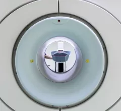
Magnetic Resonance Imaging

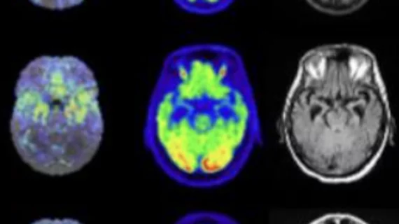
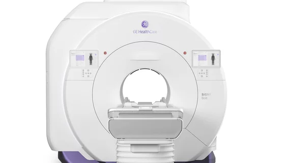
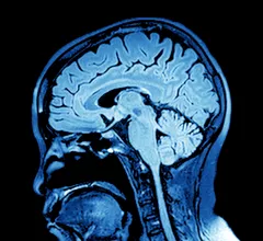
Magnetic resonance imaging (MRI) is the gold standard imaging modality for soft tissues. It produces detail cross-sectional images of soft tissue and bone anatomy, including muscles, tendons, ligaments, brain and organs, without the use of ionizing radiation. In addition to orthopedic imaging, MRI is also used for heart, brain and breast. MRI uses gadolinium contrast in many exams to highlight tissues and blood vessels, which enhances images and offers better diagnostic quality. It can also be used in conjunction with PET scans. How does MRI work? MR creates images by using powerful magnets to polarize hydrogen atoms in water (the body is made of of more than 80% water) so they face in one direction. A radiofrequency pulse is then used to ping these atoms, causing them to wobble, or resonate. The MRI coils detect this and computers can assemble images from the signals. Basic MRI scans will focus on the resonance of fat and water in two different sequences, which highlight and contrast different features in the anatomy.

Thumbnail

Whole-body MR angiography can identify early atherosclerosis at a population level

Whole-body MR angiography (MRA) is an effective screening tool for identifying early atherosclerosis in patients at low to intermediate risk for cardiovascular disease, according to a new study published by Radiology.

Example of pulmonary embolism (PE) detection on CT and confirmed using spectral CT. Image courtesy of Philips Healthcare

Researchers 'concerned' CT angiography topped MRA in follow-ups for pulmonary embolism

A recent study found whether a patient received MR angiography (MRA) instead of CT angiography (CTA) for a pulmonary embolism (PE) had little effect on the rate of follow-up chest CT or MRI one year later.

Novel MRI sensor allows for more sensitive imaging by tracking calcium

A novel calcium-based MRI sensor will allow for more sensitive brain imaging by tracking calcium ions to monitor deep neural activity, according to research out of the Massachusetts Institute of Technology.

Thumbnail

Multiparametric MRI with PI-RADSv2 misses ‘considerable number’ of prostate cancer lesions, could still provide value

The combination of multiparametric MRI and the recently revised Prostate Imaging Reporting and Data System version 2 (PI-RADSv2) misses a “considerable number” of clinically significant prostate cancer (PCA) lesions, according to a new study published in the American Journal of Roentgenology. The authors noted, however, that the high positive predictive value (PPV) might make this method useful for imaging active surveillance (AS) patients.

Thumbnail

Can convolutional neural networks accurately select MRI protocols?

Deep learning-based convolutional neural networks (CNNs) can help radiologists select musculoskeletal MRI protocols, according to a study published by the Journal of Digital Imaging.

Thumbnail

Gas vesicles act as natural contrast agents, could result in 'erasable' MRIs

Scientists at the California Institute of Technology are taking steps toward erasable MRIs by using contrast agents natural to the human body—gas vesicles.

Thumbnail

MR imaging a ‘suitable alternative’ to CT when evaluating patients for acute appendicitis

Physicians regularly turn to CT to evaluate patients who arrive at a hospital emergency department (ED) due to acute appendicitis. According to a new study published in Radiology, the diagnostic accuracy of MR imaging compares favorably to that of CT when diagnosing acute appendicitis, meaning patients can receive the same level of care without the ionizing radiation exposure.

Image reconstruction algorithm, MRI-derived heart strain values can aid prognosis in amyloidosis patients

Recent research found strain parameters taken from a cine MRI-based deformable registration algorithm (DRA) can determine the severity of amyloid buildup in the heart and may provide prognostic information on patients with light-chain (AL) amyloidosis.