Magnetic Resonance Imaging

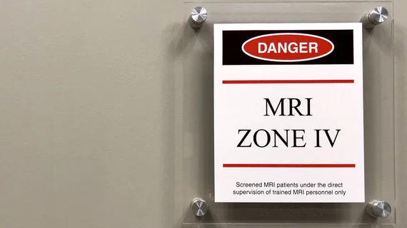
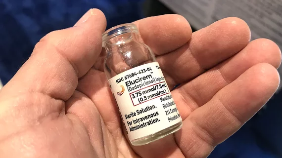
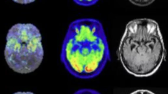
Magnetic resonance imaging (MRI) is the gold standard imaging modality for soft tissues. It produces detail cross-sectional images of soft tissue and bone anatomy, including muscles, tendons, ligaments, brain and organs, without the use of ionizing radiation. In addition to orthopedic imaging, MRI is also used for heart, brain and breast. MRI uses gadolinium contrast in many exams to highlight tissues and blood vessels, which enhances images and offers better diagnostic quality. It can also be used in conjunction with PET scans. How does MRI work? MR creates images by using powerful magnets to polarize hydrogen atoms in water (the body is made of of more than 80% water) so they face in one direction. A radiofrequency pulse is then used to ping these atoms, causing them to wobble, or resonate. The MRI coils detect this and computers can assemble images from the signals. Basic MRI scans will focus on the resonance of fat and water in two different sequences, which highlight and contrast different features in the anatomy.

Thumbnail

When CT scans are negative for patients with blunt neck injuries, follow-up MRI provides little value

When patients experience blunt neck injuries—after a car accident, for example—follow-up MRI is not necessary after a CT scan comes back negative for spine instability, according to a new study published in JAMA Surgery.

Women’s health magazine offers patient-friendly guide to MRIs

Numerous surveys have shown that patients don’t know a lot about radiology or what radiologists and technologists do. Could articles such as this patient-focused look at MRI examinations from the women’s health magazine SELF be a step toward helping patients learn more about radiology as a whole?

Thumbnail

Radiologists, meet your EMR ‘reference librarian’

Sponsored by Change Healthcare

Jeffrey B. Mendel, MD, former Chair of Radiology and Chief of Radiology Informatics at, respectively, Tufts-affiliated St. Elizabeth’s Medical Center in Boston and Harvard-affiliated Beth Israel Deaconess Medical Center in Boston, shares an anecdote.

Thumbnail

Improving workflows with McKesson Radiology advanced visualization: a 3D MR success story

McKesson

Radiologists operate in a fast-paced work environment, juggling a combination of tasks that includes interpreting studies, and calls from referring physicians.

Thumbnail

Volume and HCAHPS Scores Up, Leakage Down: St. Joseph’s Candler’s MRI Choice Made the Difference

Sponsored by Hitachi Healthcare Americas

In the MRI suite at St. Joseph’s Candler Hospital in Savannah, Ga., the numbers add up to a quantifiable success story.

Thumbnail

Vermont tests elimination of payer pre-approvals for MRIs

The Green Mountain State is in the news for more than the entry of Independent Sen. Bernie Sanders into the 2016 race for president. A pilot healthcare-delivery project there experiments with removing a rule currently requiring patients to get pre-approval from their health plans for MRIs. 

Thumbnail

Got an EMR? Get an enterprise-wide ‘clinical imaging health record’

At a time when 85% of physicians are viewing diagnostic images via electronic medical records, the opportunity is ripe to realize true enterprise imaging. More of those docs should be saying goodbye to walled-off image silos and hello to a centralized, always-open image depot.