Magnetic Resonance Imaging

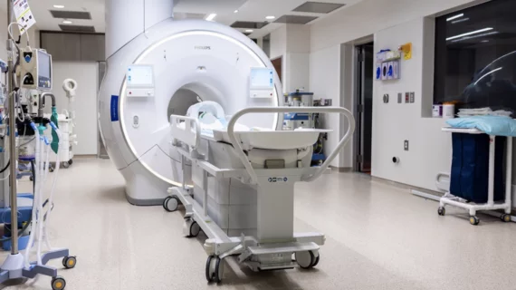
Magnetic resonance imaging (MRI) is the gold standard imaging modality for soft tissues. It produces detail cross-sectional images of soft tissue and bone anatomy, including muscles, tendons, ligaments, brain and organs, without the use of ionizing radiation. In addition to orthopedic imaging, MRI is also used for heart, brain and breast. MRI uses gadolinium contrast in many exams to highlight tissues and blood vessels, which enhances images and offers better diagnostic quality. It can also be used in conjunction with PET scans. How does MRI work? MR creates images by using powerful magnets to polarize hydrogen atoms in water (the body is made of of more than 80% water) so they face in one direction. A radiofrequency pulse is then used to ping these atoms, causing them to wobble, or resonate. The MRI coils detect this and computers can assemble images from the signals. Basic MRI scans will focus on the resonance of fat and water in two different sequences, which highlight and contrast different features in the anatomy.


Final 2014 HOPPS Rule Excludes Some Data from MR and CT Cost Center Calculations

CMS elected to keep the new separate cost centers for MR and CT in its final rule, but did drop the most flawed group of cost data from the calculations thereby reducing cuts to the 2 to 18 percent range depending on modality and procedure

Breast MRI Underused for High Cancer Risk Women

In a JAMA published study, researchers found that although breast MRI use has more than doubled in a decade, patterns of use do not reflect national cancer screening and treatment guidelines

Thumbnail

Imaging Addiction: Could PET & MR End Cocaine Abuse?

Cocaine addiction can ruin a person physically and financially, and with an estimated 1.4 million cocaine users in the U.S., thousands will become trapped by their habit. While previous research on the drug and its addictive potential were observational and subjective, imaging is reshaping how we see addiction—and how it will be treated.

SUNY Upstate: Taking the VNA Route to an Image-enriched EMR

Sponsored by FUJIFILM Healthcare Americas

Purchase a vendor-neutral archive (VNA). Design it to store DICOM and non–DICOM images. Integrate it seamlessly with electronic medical record (EMR) system. Watch its users visually enrich their patients’ medical records.

Spine MRI for Some Back Pain Added to Choosing Wisely List

North American Spine Society (NASS) adds advanced medical imaging for some back pain to list of questionable procedures kept by the American Board of Internal Medicine (ABIM) Choosing Wisely initiative

ACR Makes Official Appeal to Stop Separate CT and MR Cost Centers in 2014 HOPPS Rule

In a comment letter to CMS Administrator Marilyn Tavenner, the ACR made clear its position that the proposed separate cost centers for CT and MR in the 2014 Hospital Outpatient Prospective Payment (HOPPS) rule is a bad idea

Silva: Bad Data is Root of Problem with CT and MRI Cost Centers

Acknowledging and fixing the flawed capital cost data that CMS is using to justify proposed cuts in CT and MRI reimbursement is the responsibility of all radiology professionals writes Ezequiel Silva III, MD, in the latest issue of Radiology Business Journal

CDI Acquires The MRI Centers of New England

The Minneapolis-based outpatient imaging center network Center for Diagnostic Imaging (CDI) has expanded its regional presence in Massachusetts with the acquisition of The MRI Centers of New England