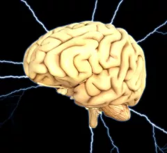
Magnetic Resonance Imaging

Magnetic resonance imaging (MRI) is the gold standard imaging modality for soft tissues. It produces detail cross-sectional images of soft tissue and bone anatomy, including muscles, tendons, ligaments, brain and organs, without the use of ionizing radiation. In addition to orthopedic imaging, MRI is also used for heart, brain and breast. MRI uses gadolinium contrast in many exams to highlight tissues and blood vessels, which enhances images and offers better diagnostic quality. It can also be used in conjunction with PET scans. How does MRI work? MR creates images by using powerful magnets to polarize hydrogen atoms in water (the body is made of of more than 80% water) so they face in one direction. A radiofrequency pulse is then used to ping these atoms, causing them to wobble, or resonate. The MRI coils detect this and computers can assemble images from the signals. Basic MRI scans will focus on the resonance of fat and water in two different sequences, which highlight and contrast different features in the anatomy.

Thumbnail

7-Tesla MRI could be a game-changer for diagnosing, treating brain tumors

A 7-Tesla-strong MRI machine could be changing the way brain tumors are diagnosed and treated, University of Southern California researchers reported this week.

Thumbnail

Machine learning model accurately predicts who would benefit most from mpMRIs

A novel machine learning model could accurately predict which men might benefit most from additional imaging before a prostate biopsy, saving patients both money and discomfort, a new study states.

High-risk breast cancer patients avoid MRIs that could improve early detection of disease

Nearly half of women at a high lifetime risk for breast cancer undergo routine mammograms at practices with onsite breast MR imaging capabilities, but MRI screenings are being vastly underused in the population, reaching just 6.6 percent of high-risk patients, a study in the Journal of Women’s Health states.

Thumbnail

Insurer approves, then denies MRI, forcing man to pay $2,340 out-of-pocket

Patients’ wishes and insurance companies’ approval don’t always line up when it comes to coverage for imaging procedures, the Philadelphia Inquirer reported this week.

Thumbnail

When CT scans are negative for patients with blunt neck injuries, follow-up MRI provides little value

When patients experience blunt neck injuries—after a car accident, for example—follow-up MRI is not necessary after a CT scan comes back negative for spine instability, according to a new study published in JAMA Surgery.

Women’s health magazine offers patient-friendly guide to MRIs

Numerous surveys have shown that patients don’t know a lot about radiology or what radiologists and technologists do. Could articles such as this patient-focused look at MRI examinations from the women’s health magazine SELF be a step toward helping patients learn more about radiology as a whole?

Thumbnail

Radiologists, meet your EMR ‘reference librarian’

Sponsored by Change Healthcare

Jeffrey B. Mendel, MD, former Chair of Radiology and Chief of Radiology Informatics at, respectively, Tufts-affiliated St. Elizabeth’s Medical Center in Boston and Harvard-affiliated Beth Israel Deaconess Medical Center in Boston, shares an anecdote.