Magnetic Resonance Imaging

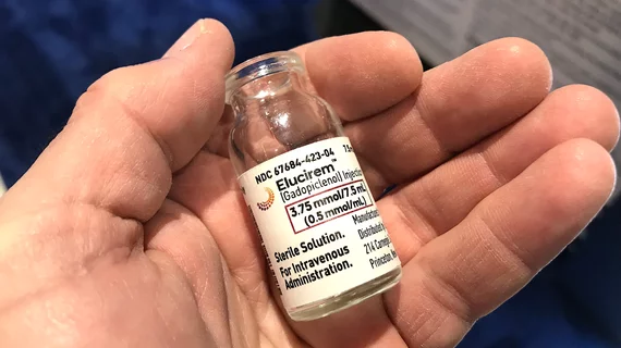
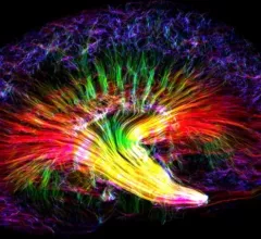
Magnetic resonance imaging (MRI) is the gold standard imaging modality for soft tissues. It produces detail cross-sectional images of soft tissue and bone anatomy, including muscles, tendons, ligaments, brain and organs, without the use of ionizing radiation. In addition to orthopedic imaging, MRI is also used for heart, brain and breast. MRI uses gadolinium contrast in many exams to highlight tissues and blood vessels, which enhances images and offers better diagnostic quality. It can also be used in conjunction with PET scans. How does MRI work? MR creates images by using powerful magnets to polarize hydrogen atoms in water (the body is made of of more than 80% water) so they face in one direction. A radiofrequency pulse is then used to ping these atoms, causing them to wobble, or resonate. The MRI coils detect this and computers can assemble images from the signals. Basic MRI scans will focus on the resonance of fat and water in two different sequences, which highlight and contrast different features in the anatomy.

GE recalls thousands of MRIs

GE is recalling almost 13,000 MRI units, as the FDA has deemed them potentially deadly.

Thumbnail

New York radiology practice replaces two MR systems with one; Significantly grows volumes

Sponsored by Hitachi Healthcare Americas

Serving the community of upstate New York’s Capital Region, Adirondack Radiology Associates (ARA) recently expanded patients’ access to state-of-the-art, high-field open bore MR imaging technology, replacing two systems, an older low-field open system and a closed 1.5T system, with the Hitachi Oasis high-field open MR at their Saratoga Imaging Center. Since the installation, ARA has experienced an increase in procedure volumes and patient satisfaction as well a reduction in costs. 

Example of a RF burn from an MRI scan where a sedated patient's identification bracelet was touching their skin during an exam. Image courtesy of RSNA. https://pubs.rsna.org/doi/10.1148/radiol.09090637

Battling Burns in MRI

While MRI is often considered one of the safer imaging modalities due to its lack of ionizing radiation, other dangers remain for patients, with thermal burns being a chief concern. 

Thumbnail

Increased Access, Dependability and Patient Satisfaction with a Fixed MRI at Star Valley CAH

Sponsored by Hitachi Healthcare Americas

Among the many requirements a hospital must meet to achieve and maintain the designation of Critical Access Hospital (CAH), the hospital must be fairly remote and furnish 24-hour emergency care services seven days a week, using either on-site or on-call staff.

National Association of Professional Women Announces Lisa Berrios-Moore, MRI Technologist, as VIP Woman of the Year

Lisa Berrios-Moore was honored by the National Association of Professional Women this week with the prestigious award and recognition as VIP Woman of the Year for her leadership in healthcare. 

Final 2014 HOPPS Rule Excludes Some Data from MR and CT Cost Center Calculations

CMS elected to keep the new separate cost centers for MR and CT in its final rule, but did drop the most flawed group of cost data from the calculations thereby reducing cuts to the 2 to 18 percent range depending on modality and procedure

Breast MRI Underused for High Cancer Risk Women

In a JAMA published study, researchers found that although breast MRI use has more than doubled in a decade, patterns of use do not reflect national cancer screening and treatment guidelines

Thumbnail

Imaging Addiction: Could PET & MR End Cocaine Abuse?

Cocaine addiction can ruin a person physically and financially, and with an estimated 1.4 million cocaine users in the U.S., thousands will become trapped by their habit. While previous research on the drug and its addictive potential were observational and subjective, imaging is reshaping how we see addiction—and how it will be treated.