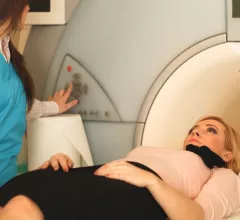
Magnetic Resonance Imaging

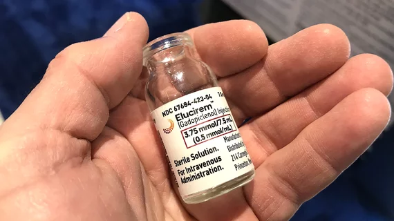
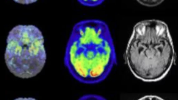
Magnetic resonance imaging (MRI) is the gold standard imaging modality for soft tissues. It produces detail cross-sectional images of soft tissue and bone anatomy, including muscles, tendons, ligaments, brain and organs, without the use of ionizing radiation. In addition to orthopedic imaging, MRI is also used for heart, brain and breast. MRI uses gadolinium contrast in many exams to highlight tissues and blood vessels, which enhances images and offers better diagnostic quality. It can also be used in conjunction with PET scans. How does MRI work? MR creates images by using powerful magnets to polarize hydrogen atoms in water (the body is made of of more than 80% water) so they face in one direction. A radiofrequency pulse is then used to ping these atoms, causing them to wobble, or resonate. The MRI coils detect this and computers can assemble images from the signals. Basic MRI scans will focus on the resonance of fat and water in two different sequences, which highlight and contrast different features in the anatomy.

MRI Offers No Added Value In RA Risk Stratification

MRI is no more cost-effective than standard risk stratification techniques in determining whether newly diagnosed rheumatoid arthritis (RA) patients will benefit from aggressive treatment is no more cost-effective than standard risk stratification techniques, according to researchers at the Yale School of Medicine in New Haven, Conn.

MR Systems Garner FDA Clearance

GE Healthcare yesterday announced FDA clearance of its Optima™ MR360 1.5T and Brivo™ MR355 1.5T magnetic resonance systems. Approximately 180 such systems have been ordered by hospitals and imaging centers worldwide.

Toshiba Launches MR System Upgrade Program

Toshiba America Medical Systems, Inc. has introduced an upgrade program for its Vantage™ 1.5T MR systems. Promoted as a cost-effective alternative to purchasing a new system, the renew ™ program offers Toshiba Vantage users a number of options throughout the entire Vantage suite, including upgrade paths to a Vantage Atlas® or a Vantage Titan™.

Musculoskeletal MR System Receives FDA Clearance

GE Healthcare late last week announced FDA clearance of its new Optima™ MR430s 1.5T scanner. Intended to deliver precise imaging of the arm, including the elbow, wrist, and hand; or the leg, including the knee, ankle, and foot, the musculoskeletal MR system delivers 70mT/m of strength and 300T/m/s of slew rate. Short echo spacing and a high signal

Soteria Imaging Services Sells Champlain Valley Open MRI, Open MRI of Lafayette

Soteria Imaging Services, Louisville, Ky., recently completed the sale of two of its imaging facilities, with Plattsburgh, N.Y.-based CPVH Medical Center acquiring Champlain Valley Open MRI, also located in Plattsburgh; and Center For Diagnostic Imaging (CDI), Burnsville, Minn., acquiring Open MRI of Lafayette, Lafayette, Ind.

Breast MRI Screening A Boon to Women With Radiation Therapy History

Breast cancer screening with MRI can detect invasive cancers missed by mammography in women who have been treated with radiation therapy to the chest for other conditions, according to a new study published online today in Radiology and slated to appear in print in the magazine’s April issue.

PET/MRI System Receives CE Marking; 510 (k) Pending Approval

Opening the door for commercial availability in Europe, The European Union (EU) has granted CE marking to Philips Healthcare’s Ingenuity TF whole body PET/MR system. Billed as the first commercially available unit of its kind, the system—which combines the molecular imaging capabilities of PET with the soft tissue contrast of MR to image the

ACR Unveils Breast MRI Accreditation Program

Sponsored by Hitachi Healthcare Americas

To enable imaging facilities to improve and maintain the quality of their breast MRI services, the ACR® has launched the Breast Magnetic Resonance Imaging Accreditation Program (BMRAP). Like other ACR accreditation programs, BMRAP involves peer-review assessment of imaging centers’ processes and equipment, along with the caliber of their images and