Magnetic Resonance Imaging

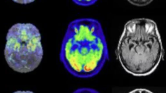
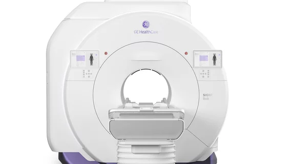
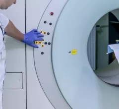
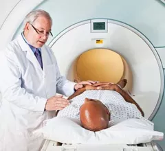
Magnetic resonance imaging (MRI) is the gold standard imaging modality for soft tissues. It produces detail cross-sectional images of soft tissue and bone anatomy, including muscles, tendons, ligaments, brain and organs, without the use of ionizing radiation. In addition to orthopedic imaging, MRI is also used for heart, brain and breast. MRI uses gadolinium contrast in many exams to highlight tissues and blood vessels, which enhances images and offers better diagnostic quality. It can also be used in conjunction with PET scans. How does MRI work? MR creates images by using powerful magnets to polarize hydrogen atoms in water (the body is made of of more than 80% water) so they face in one direction. A radiofrequency pulse is then used to ping these atoms, causing them to wobble, or resonate. The MRI coils detect this and computers can assemble images from the signals. Basic MRI scans will focus on the resonance of fat and water in two different sequences, which highlight and contrast different features in the anatomy.

Bariatric MRI: Challenges and Work-arounds

Sponsored by Hitachi Healthcare Americas

ThedaCare is Northeastern Wisconsin’s largest community-owned health system; its numerous radiology units strive to provide the highest-quality imaging to attract new referrals while simultaneously retaining hard-won existing business. Until relatively recently, however, performing MRI exams for patients weighing more than 300 pounds was a problem.

Deadline 2012: MRI Accreditation

Sponsored by Hitachi Healthcare Americas

Under the Medicare Improvements for Patients and Providers Act (MIPPA) of 2008, those sites providing the technical component of advanced imaging must have accreditation by 2012—but not all imaging centers are waiting for the deadline. A case in point is that of the Frye Care Outpatient Imaging Center, which is operated by the Frye Regional Medical

Software Allows Virtual PET/MRI Fusion

Sponsored by FUJIFILM Healthcare Americas

It was a difficult, but not uncommon, neuroradiology case: A patient suffered seizures, but had, a year before, been cleared of the possibility of epilepsy by a radiologist looking at an MRI study of the patient’s brain. The persistence of the symptoms suggested that the initial diagnosis had been incorrect, so Meng Law, MD, professor of radiology

Opening Minds to Open MRI

Sponsored by Hitachi Healthcare Americas

How do you acquire the high–field-strength open MRI unit of your dreams in a tough economy? Unless you had already budgeted a cool million before the economy tanked, it may be difficult. David Nelson, director of outpatient imaging for BayCare Health System, Clearwater, Fla, overcame his lack of comfort with open systems last year and did just that

How I Do It: Using 3D MRI to Identify High-risk Vascular Disease

Stroke and heart attack are major killers, not only in the Western world but, increasingly, throughout the developing industrial nations. The cost of these diseases is counted not only in lives lost, but in the health care costs incurred by those patients suffering the chronic manifestations of stroke and cardiac failure. While the acute and more

ACR Unveils a Modular MRI Accreditation Process

Sponsored by Hitachi Healthcare Americas

The ACR is set to launch a new modular MRI accreditation program designed to meet the specific practice patterns of individual facilities. Sometime this month, the current general MRI accreditation program will transition to the new program—which includes brain, spine, musculoskeletal, body, MR angiography, and cardiac modules—opening the door to

Coronary MR Angiography: Can It Compete With Multidetector CT?

Just five years ago, the thought of cardiac cross-sectional imaging turned immediately toward the potential of MRI for evaluation of the heart. Equipment manufacturers provided new MRI scanners with fast gradients for this purpose and developed new pulse sequences. Advanced 3D workstations provided new software for analysis of cardiac function, and

Look, No Hands: Reports Go Directly to Referrer EMRs

John Griffith, CIO of Epic Imaging, Portland, Ore, says, “They asked us for it, and we had to come up with a solution.” The radiology practice provides imaging for close to 2,000 physician referrers; what they wanted was electronically delivered radiology reports sent to their practices not via fax, nor even over the Web, where they had to reside