Computed Tomography

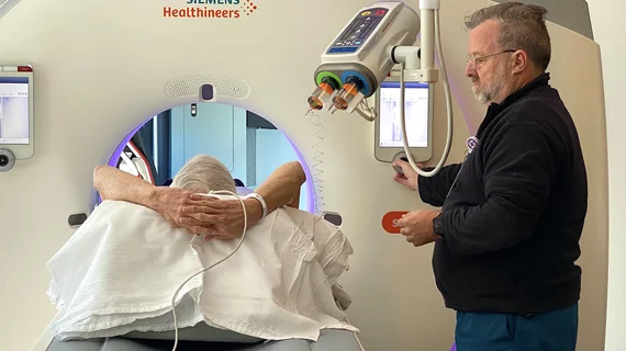
Computed tomography (CT) is a fast and accurate imaging modality often used in emergency settings and trauma imaging. CT scans, with or without (or both) iodinated contrast are frequently used to image the brain, chest, abdomen and pelvis, but also have post-imaging reconstructive capabilities for detailed orthopedic imaging. It is now a standard imaging modality in emergency rooms to quickly assess patients. CT uses a series of X-ray images shot as the gantry rotates around the patient. Computer technology assembles these into into a dataset volume than can be slices on any access, or advanced visualization software can extract specific parts of the anatomy for study. Find more content specific to cardiac CT.

Emergency overreads change care for 92% of patients with discrepant outside reports

When emergency radiologists overread interpretations of critically ill transfer patients, they discover discrepancies in fewer than 15% of cases. However, more than 90% of these second opinions produce a change in patient care or follow-up.

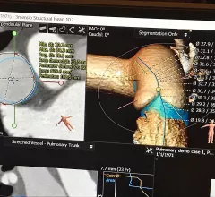
Vascular CT beyond the coronaries includes imaging for aortic aneurism, aortic dissection, SCAD and fibromuscular dysplasia. #SCCT #SCCT2022 #SCAD #yescct

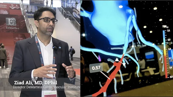
VIDEO: Vascular imaging applications for CT beyond the heart

Society of Cardiovascular Computed Tomography (SCCT) President Brian Ghoshhajra, MD, explains the vascular imaging sessions at SCCT 2022 that went beyond the usual coronaries and structural heart evaluations.

Faster CT translates to fewer sedated children

Upgrading to dual-source, dual-energy CT machines cut average pediatric scan times from around 12 seconds to three seconds or less at two sites of an academic emergency department.

FDA greenlights ortho robot, automated MRI brain modeling

FDA approvals arrive for ClearPoint Neuro and MicroPort Navibot. 

coronary CTA

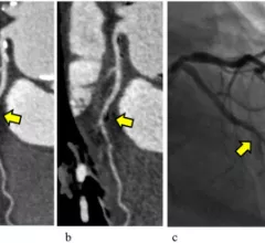
Deep learning, subtraction technique ideal for evaluating stent re-stenosis on coronary CTA

Detecting in-stent restenosis via coronary CTA with hybrid iterative reconstruction has historically been an exercise in avoiding false positives.

Thumbnail

These CT findings increase risk of thromboembolic events for patients with COVID pneumonia

Authors of the new paper noted that radiologists should be highly suspicious of pulmonary embolism and DVT when they encounter these findings on chest CT exams of COVID patients.

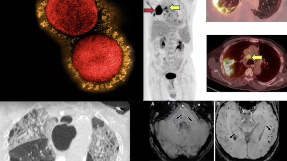
COVID-19 medical imaging examples of various clinical presentations. SARS-CoV-2 clinical imaging presentations.

PHOTO GALLERY: What does COVID-19 look like on medical imaging?

This image gallery shows what the various clinical presentations associated with the COVID-19 virus that have been documented during the coronavirus pandemic.

What works—and what doesn’t—for chipping away at CT overutilization in the ED

The presence of any or all of four factors can help ensure appropriate CT utilization in emergency settings: established diagnostic pathways, alternative test availability, involvement of specialists and feedback from referrers.