Computed Tomography

Computed tomography (CT) is a fast and accurate imaging modality often used in emergency settings and trauma imaging. CT scans, with or without (or both) iodinated contrast are frequently used to image the brain, chest, abdomen and pelvis, but also have post-imaging reconstructive capabilities for detailed orthopedic imaging. It is now a standard imaging modality in emergency rooms to quickly assess patients. CT uses a series of X-ray images shot as the gantry rotates around the patient. Computer technology assembles these into into a dataset volume than can be slices on any access, or advanced visualization software can extract specific parts of the anatomy for study. Find more content specific to cardiac CT.

Thumbnail

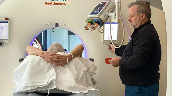
Digitized informed consent increases detection of critical 'red flags' prior to imaging exams

Digitized informed patient consent prior to contrast-enhanced CT exams has significant advantages over traditional paper consent forms, but it also has the unfortunate effect of leaving some of the most vulnerable patients behind.  

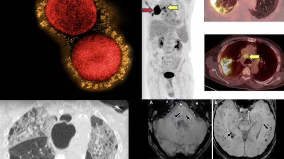
long covid lung CT

More than half of long COVID patients have abnormalities on chest imaging

Nodular patterns were the most commonly visualized abnormality, although several others were observed.

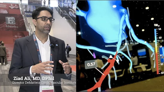
Left, HeartFlow's RoadMap analysis enables cardiac CT readers to identify stenoses in the major coronary arteries. The AI provides visualization and quantification of the location and severity of anatomic narrowings. Right image, HeartFlow's Plaque Analysis AI algorithm automates assessment of coronary plaque characteristics and volume on CCTA exams to greatly reduce the time it takes to manually assess and quantify these features.

HeartFlow gains FDA clearance for 2 new AI-powered imaging assessments

The solutions, Plaque Analysis and RoadMap Analysis, both use coronary CT angiography to provide clinicians with a noninvasive look at patients who present with coronary artery disease and face a heightened myocardial infarction risk.

Thumbnail

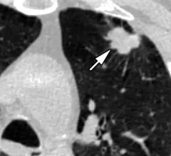
CRC patients with these clinical characteristics need more frequent post-op chest imaging

These patients are at greatest risk of developing lung metastases within three months of surgery.

Total body PET/CT scans may offer benefits for evaluating arthritis

Low-dose scans showed high agreement with joint-by-joint rheumatological evaluations. 


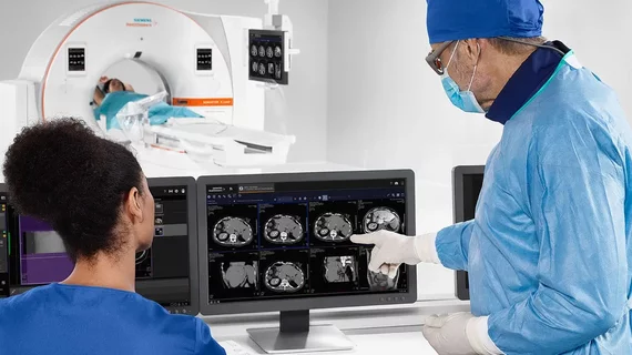
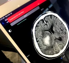
AI system boosts intracranial hemorrhage detection

“This study implies that future clinical workflows may see AI be used in an adjunct capacity to improve interpretations of CT scans by helping call radiologists' attention to findings that may be overlooked.” 

2 imaging technologies cleared for respective markets

The FDA has approved a CT guidance system for interventional radiologists performing percutaneous procedures with robotics. And the European Union has greenlit a family of mobile CT scanners.

VIDEO: Cardiac CT as a front-line chest pain exam in the ED

Harold Litt, MD, explains the use of cardiac CT as a primary imaging exam for chest pain at his emergency department over the past decade.