Computed Tomography

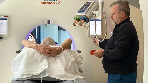
Computed tomography (CT) is a fast and accurate imaging modality often used in emergency settings and trauma imaging. CT scans, with or without (or both) iodinated contrast are frequently used to image the brain, chest, abdomen and pelvis, but also have post-imaging reconstructive capabilities for detailed orthopedic imaging. It is now a standard imaging modality in emergency rooms to quickly assess patients. CT uses a series of X-ray images shot as the gantry rotates around the patient. Computer technology assembles these into into a dataset volume than can be slices on any access, or advanced visualization software can extract specific parts of the anatomy for study. Find more content specific to cardiac CT.

Thumbnail

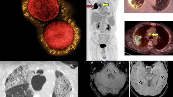
New lesion measurement assesses treatment responses more accurately than RECIST

Researchers believe the new method of assessing treatment responses could pave the way for developing new cancer therapeutics.

Thumbnail

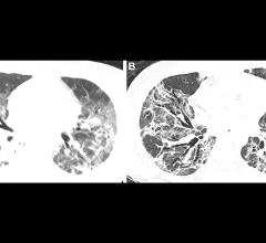
Transplanted lungs react to COVID in a distinctive way

Clinicians treating COVID-19 patients who have transplanted lungs and lower airway infection should order molecular testing in addition to, or regardless of, imaging findings.

CT imaging showing the congenital heart defect of left heart hypoplastic syndrome.

VIDEO: Congenital heart imaging with cardiac CT

As the population of patients with adult congenital heart disease grows, they are presenting to adult cardiology clinics and being imaged with CT. Many also do not have access or cannot be imaged by MRI, said Renee Bullock-Palmer, MD.

Representative cases showing pneumonia extents and patterns on chest X-ray (CXRs) and CT images. (E and F) A 36-year-old male with no history of vaccination for COVID- 19. The patient had no history of comorbidity. Axial chest CT image obtained on the same day showing unilateral ground-glass opacity with a nonrounded morphology and non-peripheral distribution in the left upper lobe (arrows). RSNA Image. COVID on X-ray, CT scan. What does COVID look like in medical imaging? Example of COVID imaging.

Vaccinated vs unvaccinated, Delta vs Omicron—how do these factors impact clinical and imaging features?

Vaccination rates likely had a role in reducing disease severity during Omicron, a new paper published in Radiology suggests.

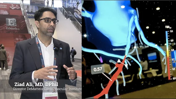
New recommendations for cardiac CT in cardio-oncology imaging

The statement was written to fill gaps in recommendations from prior consensus statements and guidelines in regards to the use of CCT in cardio-oncology, including use of calcium scoring and ruling out coronary disease when cardiac function is impaired.

Thumbnail

Experts concerned with radiation doses in young women call for stricter ordering criteria for CT exams

Experts found that compared to the average effective doses of organs, breast doses were higher, representing increased risks of breast cancer for women. 

Long and longer: Imaging wait times in Canada

Prior to the arrival of COVID-19 in early 2020, Canadian patients waited nearly three months for an MRI and more than 11 weeks for a CT. Things have only gotten worse since then.

Thumbnail

'Radiologic serendipity' a common trigger event that ends in thyroid surgery in asymptomatic patients

In this cohort, experts found that just 34% of surgeries were performed on symptomatic patients, which shifted their focus to other potential modes of detection.