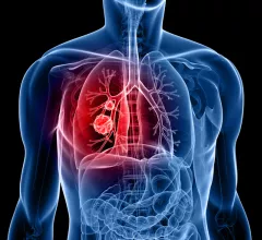
Computed Tomography

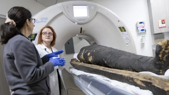
Computed tomography (CT) is a fast and accurate imaging modality often used in emergency settings and trauma imaging. CT scans, with or without (or both) iodinated contrast are frequently used to image the brain, chest, abdomen and pelvis, but also have post-imaging reconstructive capabilities for detailed orthopedic imaging. It is now a standard imaging modality in emergency rooms to quickly assess patients. CT uses a series of X-ray images shot as the gantry rotates around the patient. Computer technology assembles these into into a dataset volume than can be slices on any access, or advanced visualization software can extract specific parts of the anatomy for study. Find more content specific to cardiac CT.

Some neurovascular imaging studies are overutilized in stroke triage

Triaging patients with stroke-like symptoms using MRI with diffusion-weighted imaging (DWI) could reduce the cost burden associated with unnecessary neurovascular imaging procedures. 

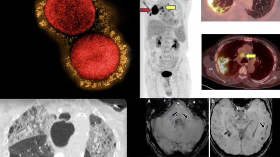
Detailed MRI reveals pulmonary vascular abnormalities in COVID long-haulers

Researchers had 40 participants inhale polarized xenon gas in order to conduct 129Xe MRI exams for the study.

Philips picks partner for multispecialty management of lung cancer

An imaging OEM is teaming with a multi-omics diagnostics company to offer lung-cancer care teams lab data alongside radiologic findings and clinical histories.

Thumbnail

'One-stop-shop' CT protocol saves time, reduces radiation needed for acute stroke imaging

The protocol does not come at the expense of deteriorated image quality, according to work published recently in the European Journal of Radiology.

enlarged parathyroid gland

Routine CT scans offer opportunistic assessments of the parathyroid gland

Such assessments could “decrease existing diagnosis gaps,” according to a new study published in Academic Radiology

Thumbnail

Radiologists develop AI to flag artifacts on CT pulmonary angiography

The capability could allow immediate alerting of CT technologists, who would adjust scan protocols or re-scan patients to optimize image quality prior to physician interpretation.

Thumbnail

How do Omicron and Delta compare on chest imaging?

Researchers found that 37% of patients with Omicron had CT scans that were considered normal compared to 15% with Delta. 

Arl Van Moore, MD, American College of Radiology (ACR) delegate to the American Medical Association (AMA) House of Delegates, former ACR president, chairman of the ACR Board and former CEO of Strategic Radiology, discusses two radiology related policy decisions at the AMA 2022 meeting. These included a AMA policy on the iodine contrast shortage and AUC CDS. #AMA #AMA22 #AMAmtg #AMA175 #ACR

VIDEO: Radiology takeaways from the 2022 AMA meeting

Arl Van Moore, MD, American College of Radiology (ACR) delegate to the American Medical Association House of Delegates, former ACR president, chairman of the ACR Board and former CEO of Strategic Radiology, discusses radiology-related policy decisions at the AMA 2022 meeting.