Computed Tomography

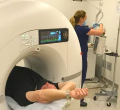
Computed tomography (CT) is a fast and accurate imaging modality often used in emergency settings and trauma imaging. CT scans, with or without (or both) iodinated contrast are frequently used to image the brain, chest, abdomen and pelvis, but also have post-imaging reconstructive capabilities for detailed orthopedic imaging. It is now a standard imaging modality in emergency rooms to quickly assess patients. CT uses a series of X-ray images shot as the gantry rotates around the patient. Computer technology assembles these into into a dataset volume than can be slices on any access, or advanced visualization software can extract specific parts of the anatomy for study. Find more content specific to cardiac CT.

Thumbnail

Experts concerned with radiation doses in young women call for stricter ordering criteria for CT exams

Experts found that compared to the average effective doses of organs, breast doses were higher, representing increased risks of breast cancer for women. 

Long and longer: Imaging wait times in Canada

Prior to the arrival of COVID-19 in early 2020, Canadian patients waited nearly three months for an MRI and more than 11 weeks for a CT. Things have only gotten worse since then.

Thumbnail

'Radiologic serendipity' a common trigger event that ends in thyroid surgery in asymptomatic patients

In this cohort, experts found that just 34% of surgeries were performed on symptomatic patients, which shifted their focus to other potential modes of detection.

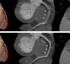
Examples of structural heart transcatheter valve replacement procedure planning CT scans and post procedure followup for TMVR and TAVR.

VIDEO: CT imaging for TAVR and TMVR structural heart interventions

Joao Cavalcante, MD, director, cardiac MRI and structural CT labs, Minneapolis Heart Institute, discusses the use of cardiac CT imaging to plan and guide structural heart procedures. 

Thumbnail

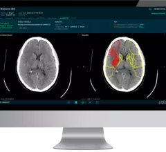
AI identifies pancreatic cancer frequently missed on CT

Specifically, the computer-aided detection (CAD) tool is capable of identifying lesions that are less than 2 cm.

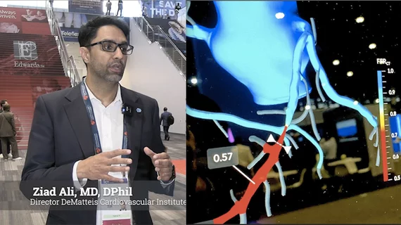
VIDEO: Gender differences in women with cardiovascular disease and implications for imagers

Erin D. Michos, MD, co-editor in chief of the American Journal of Preventive Cardiology, discusses gender differences in heart disease presentations.

AI for assessing bone fragility gets breakthrough tag

Orthopedic cone-beam CT supplier CurveBeam AI has received the FDA’s breakthrough device designation for software that computes risk of fracture in patients with osteopenia.

Photon-counting CT/AI combo improves multiple myeloma detection

When combined with artificial intelligence-based noise reduction techniques, new photon-counting CT technology can increase the detection of bone disease while also decreasing radiation exposure.