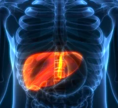
Computed Tomography

Computed tomography (CT) is a fast and accurate imaging modality often used in emergency settings and trauma imaging. CT scans, with or without (or both) iodinated contrast are frequently used to image the brain, chest, abdomen and pelvis, but also have post-imaging reconstructive capabilities for detailed orthopedic imaging. It is now a standard imaging modality in emergency rooms to quickly assess patients. CT uses a series of X-ray images shot as the gantry rotates around the patient. Computer technology assembles these into into a dataset volume than can be slices on any access, or advanced visualization software can extract specific parts of the anatomy for study. Find more content specific to cardiac CT.

Thumbnail

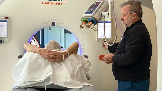
3D camera guidance meaningfully aids patient positioning, dose reduction

Regardless of a patient’s body mass index, using a scanner-integrated 3D camera for situating CT patients on the table can optimize not only patient positioning but also radiation dose. 

#CTA #acuteischemicstroke #AIS #radiomics

CTA-based radiomics can reliably estimate time since stroke onset

Radiomics features could be especially useful for determining TSS in scenarios where clinical data is lacking or unreliable, such as when patients have altered mental status.

#CTA #photoncountingCT #aortaCT

Photon counting cuts CTA contrast dosage 25%

Not only does the low-volume contrast protocol preserve supplies, it also protects patients who might be vulnerable to adverse reactions and/or side effects from contrast use. 

Rising trauma CT utilization flagged over costs, incidental findings

The increase has been especially evident in single-encounter chest/abdominopelvic scans, not least those performed for minor injuries.

Thumbnail

A 'clear lack of adherence' in imaging guidelines is impacting lung cancer survival rates

A study published in the January issue of the Journal of Nuclear Medicine reveals that nearly half of Medicare patients with NSCLC in the United States do not complete recommended FDG PET/CT scans prior to treatment.

Example of artificial intelligence generated measurements to quantify the size of a lung cancer nodule during a followup CT scan to see if the lesion is regressing with treatment. This type of automation can aid radiologists by doing the tedious, time consuming work. Photo by Dave Fornell

8 trends in radiology technology to watch in 2023

Here is a list of some key trends in radiology technology from our editors based on our coverage of the radiology market.

New type of PET/CT scan could change how hypertension is diagnosed and treated

The 10-minute scan can “light up” aldosterone-producing nodules in the adrenal glands following an injection of metomidate—a radioactive dye that binds specifically to aldosterone-producing nodules.

cerebrovascular injury #traumaimaging #vascularimaging #vascularinjury

New research discourages use of advanced vascular imaging in trauma patients

Experts argue that the overall incidence of blunt cerebrovascular injury is very low and that symptomatic vascular injuries in these cases are even lower.