Computed Tomography

Computed tomography (CT) is a fast and accurate imaging modality often used in emergency settings and trauma imaging. CT scans, with or without (or both) iodinated contrast are frequently used to image the brain, chest, abdomen and pelvis, but also have post-imaging reconstructive capabilities for detailed orthopedic imaging. It is now a standard imaging modality in emergency rooms to quickly assess patients. CT uses a series of X-ray images shot as the gantry rotates around the patient. Computer technology assembles these into into a dataset volume than can be slices on any access, or advanced visualization software can extract specific parts of the anatomy for study. Find more content specific to cardiac CT.

Thumbnail

Why do many of America's most at-risk veterans decline annual CT lung cancer screening?

The new analysis included 43,000 eligible U.S. veterans, 32% of whom were observed to have declined annual CT screening for lung cancer.

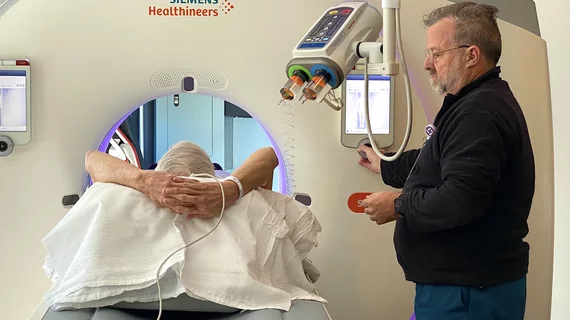
woolly mammoth tusk CT

A mammoth accomplishment: CT captures images of an entire ancient tusk

Prior attempts at imaging large fossils such as mammoth tusks failed to capture the full artifact with just one scan, instead requiring multiple partial scans that were subsequently pieced together.

Example of an artificial intelligence (AI) app store on the Sectra website, where Sectra PACS users can select the AI algorithms they want that are already integrated into the Sectra System. Other vendors have followed a similar approach to AI developed by many smaller vendors they partner with.

VIDEO: Development of AI app stores to enable easier access

Keith J. Dreyer, DO, PhD, FACR, American College of Radiology (ACR) Data Science Institute Chief Science Officer, explains how radiology vendors have developed AI app stores to make it easier to access new FDA cleared AI algorithms.
 

Keith J. Dreyer, DO, PhD, FACR, American College of Radiology (ACR) Data Science Institute Chief Science Officer, explains artificial intelligence (AI) for radiology. Dreyer also holds the positions of vice chairman of radiology at Massachusetts General Hospital, chief data science and information officer for the departments of radiology for both Massachusetts General Hospital and Brigham and Women's Hospital.

VIDEO: Where will radiology AI be in 5 years?

Keith J. Dreyer, DO, PhD, FACR, American College of Radiology (ACR) Data Science Institute Chief Science Officer, explains 5 developments to watch for in radiology artificial intelligence (AI).

Emergency overreads change care for 92% of patients with discrepant outside reports

When emergency radiologists overread interpretations of critically ill transfer patients, they discover discrepancies in fewer than 15% of cases. However, more than 90% of these second opinions produce a change in patient care or follow-up.

Vascular CT beyond the coronaries includes imaging for aortic aneurism, aortic dissection, SCAD and fibromuscular dysplasia. #SCCT #SCCT2022 #SCAD #yescct

VIDEO: Vascular imaging applications for CT beyond the heart

Society of Cardiovascular Computed Tomography (SCCT) President Brian Ghoshhajra, MD, explains the vascular imaging sessions at SCCT 2022 that went beyond the usual coronaries and structural heart evaluations.

Faster CT translates to fewer sedated children

Upgrading to dual-source, dual-energy CT machines cut average pediatric scan times from around 12 seconds to three seconds or less at two sites of an academic emergency department.

FDA greenlights ortho robot, automated MRI brain modeling

FDA approvals arrive for ClearPoint Neuro and MicroPort Navibot.