Computed Tomography

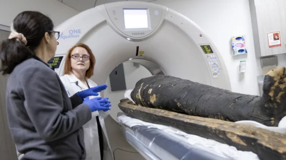
Computed tomography (CT) is a fast and accurate imaging modality often used in emergency settings and trauma imaging. CT scans, with or without (or both) iodinated contrast are frequently used to image the brain, chest, abdomen and pelvis, but also have post-imaging reconstructive capabilities for detailed orthopedic imaging. It is now a standard imaging modality in emergency rooms to quickly assess patients. CT uses a series of X-ray images shot as the gantry rotates around the patient. Computer technology assembles these into into a dataset volume than can be slices on any access, or advanced visualization software can extract specific parts of the anatomy for study. Find more content specific to cardiac CT.

long covid lung CT

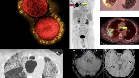
Experts describe how long-term lung abnormalities of COVID patients present on imaging

Up to one-third of patients hospitalized with COVID-19 pneumonia have abnormalities on chest CT 12 months after infection.

Ischemic stroke shown in CT scans. Image courtesy of RSNA

VIDEO: AI for stroke detection on CT imaging

Bibb Allen, MD, FACR, chief medical officer of the American College of Radiology (ACR) Data Science Institute, explains the trend of using AI for the automated detection of stroke on computed tomography (CT) imaging and the need to include radiologists on the stroke care team.

lung cancer pulmonary nodule

How effective are guidelines for managing incidental pulmonary nodules spotted on CT?

The authors of the study hypothesized that they would find malalignments due to the nature of how the 2017 Fleischner Society guidelines, in that they do not pertain to incidentally detected nodules.

pancreas lesions on CT

DL model's pancreatic lesion detection in line with that of board-certified radiologists

“Our approach has the potential to facilitate timely diagnoses and management of pancreatic lesions encountered in routine clinical practice,” experts involved in the study said.

Thumbnail

Emergency trunk CT rises sharply even for minor, moderate trauma

Almost 10 of every 1,000 patients evaluated for trauma injuries in 2018 received CT scans of the chest, abdomen and pelvis to supply a diagnostic view of, in essence, the entire torso.

Fully automated CT body composition analysis predicts survival for CRC patients

A fully automated body composition analysis derived from CT imaging can be a valuable pretreatment tool for patients with colorectal cancer. 

non-small cell lung cancer tumor segmentation

Algorithm reduces NSCLC tumor segmentation times by 65%

In a close collaboration with radiation oncologists, experts trained their model on the CT lung images of 787 patients and tested it on the scans of more than 1,300 patients from external datasets.

Thumbnail

5 imaging modalities have changed the war on cancer over the past 50 years. How many can you name?

Two branch chiefs with the cancer imaging program at NIH’s National Cancer Institute have produced a written commemoration to mark the golden anniversary of 1971’s National Cancer Act.