Computed Tomography

Computed tomography (CT) is a fast and accurate imaging modality often used in emergency settings and trauma imaging. CT scans, with or without (or both) iodinated contrast are frequently used to image the brain, chest, abdomen and pelvis, but also have post-imaging reconstructive capabilities for detailed orthopedic imaging. It is now a standard imaging modality in emergency rooms to quickly assess patients. CT uses a series of X-ray images shot as the gantry rotates around the patient. Computer technology assembles these into into a dataset volume than can be slices on any access, or advanced visualization software can extract specific parts of the anatomy for study. Find more content specific to cardiac CT.

Ed Nicol, MD, MBA, FSCCT, consultant cardiologist, honorary senior clinical lecturer, Kings College London, and president-elect of the Society of Cardiovascular Computed Tomography (SCCT), discusses what he sees as the big technology advances in cardiac CT. #SCCT #SCCT22 #RSNA22

VIDEO: New cardiac CT advances to watch

Ed Nicol, MD, MBA, president-elect of the Society of Cardiovascular Computed Tomography, discusses what he sees as the big technology advances in cardiac CT.

Leslee Shaw, PhD, MSCCT, FACC, MASNC, FAHA, director of the Blavatnik Family Women's Health Research Institute, a professor of medicine at the Icahn School of Medicine at Mount Sinai, and a former president of both SCCT and ASNC. She explains the sex differences in cardiovascular imaging presentations in women versus men.

VIDEO: CT can play a role in identifying women's differences in cardiovascular presentations

Leslee Shaw, PhD, director of the Blavatnik Family Women's Health Research Institute, a professor of medicine at the Icahn School of Medicine at Mount Sinai, and a former president of both SCCT and ASNC, explains the sex differences in cardiovascular imaging presentations in women versus men. 

Ricardo Cury, MD, MBA, MSCCT, chairman of radiology, direct of cardiac imaging, Baptist Health South Florida and Miami Cardiac and Vascular Institute, discusses the new CAD-RADS 2.0 cardiac imaging reporting criteria at the 2022 SCCT meeting. Interview with Radiology Business Editor Dave Fornell.

VIDEO: What is new with CAD-RADS 2.0 cardiac imaging reporting?

Ricardo Cury, MD, chairman of radiology and director of cardiac imaging, Baptist Health South Florida and Miami Cardiac and Vascular Institute, discusses the new CAD-RADS 2.0 cardiac imaging reporting criteria.

$75M malpractice verdict splits fault between ER doctor, radiologist

A jury in Georgia has pinned 60% of the blame for a stroke patient’s permanent whole-body paralysis on an ER physician and 40% on a radiologist—while clearing all other clinicians who had a hand in the catastrophic episode of care.

AI-generated coronary tree from a patient's CT scan showing a color code of areas of interest for plaque burden from the Cleerly software shown at SCCT 2022.

VIDEO: The role of AI in cardiac imaging

Ed Nicol, MD, president-elect of the Society of Cardiovascular Computed Tomography, provided us with an exclusive look at how AI is expected to change cardiac imaging.

Triplet pregnancy fetus in fetu: A rare case report

While FIF is an extremely rare condition—occurring in about one in 500,000 births—it is even more rare in triplet pregnancies.

Thumbnail

Digitized informed consent increases detection of critical 'red flags' prior to imaging exams

Digitized informed patient consent prior to contrast-enhanced CT exams has significant advantages over traditional paper consent forms, but it also has the unfortunate effect of leaving some of the most vulnerable patients behind.  

long covid lung CT

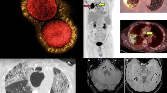
More than half of long COVID patients have abnormalities on chest imaging

Nodular patterns were the most commonly visualized abnormality, although several others were observed.