Breast Imaging

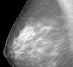
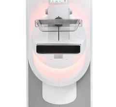
Breast imaging includes imaging modalities used for breast cancer screenings and planning therapy once cancer is detected. Mammography is the primary modality used. Mammogram technology is moving from 2D full-field digital mammography (FFDM) to breast tomosynthesis, or 3D mammography, which helps reduce false positive exams by allowing radiologists to look through the layers of tissue. Overlapping areas of dense breast tissue on 2D mammograms appear similar to cancers and 3D tomo helps determine if suspect areas are cancer or not. About 50% of women have dense breast tissue, which appears white on mammograms, the same as cancers, making diagnosis difficult. Radiologists use the Breast Imaging Reporting and Data System (BI-RADS) scoring system to define the density of breast tissue. Many states now require patients to be notified if they have dense breasts so they understand their mammograms might be suboptimal and they should use supplemental imaging that can see through the dense areas. This includes tomosythesis, breast ultrasound, automated breast ultrasound (ABUS), breast MRI, contrast enhanced mammography and nuclear imaging, including positron emission mammography (PEM).


breast radiologist breast cancer mammography

Ultrasound combined with MRI is better able to identify cancerous lesions in breast tissue

Research out of China found that the pairing proved to be more accurate in identifying cancer in nonmass-like lesions.

breast cancer month ribbon

Patients struggling with addiction, mental health are less likely to be screened for breast cancer

Researchers have shed light on the relationship between breast cancer screening and sociodemographic factors.

Paige Breast Lymph Node Screenshot

FDA grants Paige ‘Breakthrough’ status for breast cancer detection

Paige Lymph Node uses AI to assist pathologists in detecting breast cancer metastases.

breast cancer month ribbon

Most women fail to recognize signs of breast cancer, a third are confused by screening guidelines

Results of a survey from Ohio State University show the public needs a lot more education on breast cancer facts.

An RSNA attendee undergoes an MRI brain scan on the expo floor using the Hyperfine Swoop head MRI system. It is self-shields with a low field 0.064 T. It uses a standard wall power outlet and can be wheeled through a standard 34-inch wide door frame. It weighs 1,400 pounds. Imaging sequences include T1, T2, FLAIR, and DWI (with ADC map) and its operational controls are all directed on an iPad interface. #RSNA #RSNA22

FDA clears new artificial intelligence capabilities for portable MRI scanner

This is the eighth clearance in the past three years for Hyperfine's Swoop system, company officials said Monday. 

breast radiologist breast cancer mammography

Survey says most women skip their recommended mammogram

Women cite a lack of time as the primary reason for not scheduling a mammogram, according to a survey from one large health system.

How MRI “fingerprinting” could help personalize breast cancer treatment

Researchers from Case Western Reserve University are commissioning a study to determine if a novel MRI technique can predict the effectiveness of chemotherapy in treating breast cancer.

breast cancer mammography mammogram

AI proves a qualified second reader for screening mammography

According to one radiologist, the continued success of AI could make second reads for screening mammograms a thing of the past.