Molecular Imaging

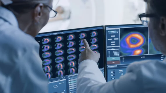
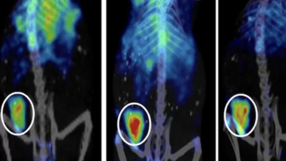
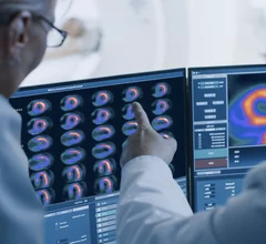
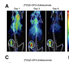
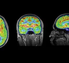
Molecular imaging (also called nuclear medicine or nuclear imaging) can image the function of cells inside the body at the molecular level. This includes the imaging modalities of positron emission computed tomography (PET) and single photon emission computed tomography (SPECT) imaging. How does PET and SPECT imaging work? Small amounts of radioactive material (radiopharmaceuticals) injected into a patient. These can use sugars or chemical traits to bond to specific cells. The radioactive material is taken up by cells that consume the sugars. The radiation emitted from inside the body is detected by photon detectors outside the body. Computers take the data to assemble images of the radiation emissions. Nuclear images may appear fuzzy or ghostly rather than the sharper resolution from MRI and CT.  But, it provides metabolic information at a cellular level, showing if there are defects in the function of the heart, areas of very high metabolic activity associated with cancer cells, or areas of inflammation, data not available from other modalities. These noninvasive imaging exams are used to diagnose cancer, heart disease, Alzheimer’s and Parkinson’s disease, bone disorders and other disorders. 

NIH grants to fund study of molecular imaging in stroke care

NIH is hopeful the research will enable doctors to detect signs of stroke damage earlier, opening the door for them to initiate treatments in a timelier manner.

Late-stage prostate cancer diagnoses on the rise

Experts say these new findings highlight the need for more effective screening initiatives among men who may face access barriers. 

Pharos PET scanner earns FDA clearance

Compact multifunctional PET scanner receives clearance in the US

The system was built with patients in mind, and its compact design has the potential to expand access to the modality where it may have previously been lacking. 

Fluorescent contrast agent helps diagnose basal cell carcinoma.

Fluorescent contrast agent noninvasively identifies common skin cancer

When used topically, the agent penetrates the skin and generates a fluorescent signal under the guidance of a fluorescent confocal microscope.

Dual-mode radiotracer helps surgeons 'hear' invisible cancer lesions

Surgeons can 'hear' cancerous lesions with help of new dual-mode radiotracer

When used during surgery, it can help providers identify cancerous lesions that are not in their immediate vision. 

Molecular Imaging PET and SPECT sign in GE booth ACC23.

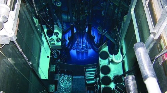
Shine working toward U.S. production of Mo-99 for SPECT imaging

Greg Piefer, PhD, chief executive and founder of SHINE, a Wisconsin-based radiopharmaceutical company that is building the world’s largest medical isotope production facility in Janesville, Wisconsin, explains progress on the company's fusion reactor that will become the first U.S. commercial supplier of molybdenum-99 in decades.

PSMA PET/CT scans can identify prostate cancer patients most likely to benefit from salvage radiotherapy

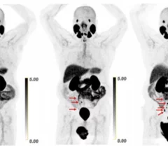
A new study is the first to show that men with biochemically recurrent prostate cancer who undergo PSMA PET/CT prior to salvage therapy have improved survival rates. 

prostate PSMA

New formulation of popular PSMA imaging agent could up availability by 50%

The U.S. Food and Drug Administration has accepted a New Drug Application for a new formulation of a commercially available PSMA imaging agent.