Breast Imaging

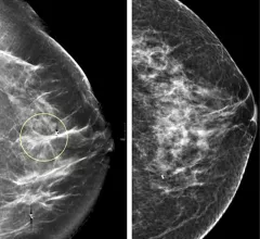
Breast imaging includes imaging modalities used for breast cancer screenings and planning therapy once cancer is detected. Mammography is the primary modality used. Mammogram technology is moving from 2D full-field digital mammography (FFDM) to breast tomosynthesis, or 3D mammography, which helps reduce false positive exams by allowing radiologists to look through the layers of tissue. Overlapping areas of dense breast tissue on 2D mammograms appear similar to cancers and 3D tomo helps determine if suspect areas are cancer or not. About 50% of women have dense breast tissue, which appears white on mammograms, the same as cancers, making diagnosis difficult. Radiologists use the Breast Imaging Reporting and Data System (BI-RADS) scoring system to define the density of breast tissue. Many states now require patients to be notified if they have dense breasts so they understand their mammograms might be suboptimal and they should use supplemental imaging that can see through the dense areas. This includes tomosythesis, breast ultrasound, automated breast ultrasound (ABUS), breast MRI, contrast enhanced mammography and nuclear imaging, including positron emission mammography (PEM).

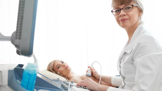
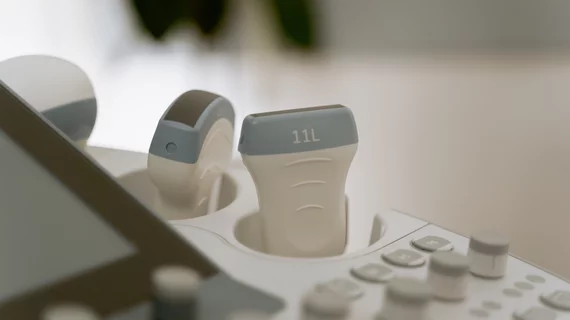
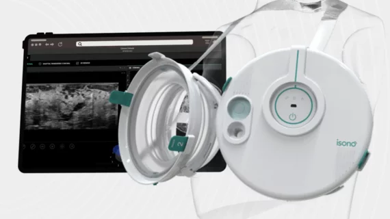
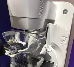
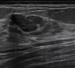
Automated breast ultrasound (ABUS) allows for reproducible breast imaging without variation based on which sonographer performs the exam. It also can help centers were they are short on qualified breast sonographers.  Breast ultrasound can help identify cancers, or benign cysts, even in women with very dense breast tissue. At the GE Healthcare booth at RSNA.

Automated ultrasound excels for women with dense breasts

Could automated breast ultrasound replace handheld ultrasound?

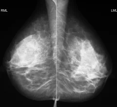
Example of the four types of breast tissue density. The density of fibroglandular tissue inside the breast impacts the ability to easily see cancers. Cancers are very easy to spot in fatty breasts, but are almost impossible to find in extremely dense breasts. These examples show craniocaudal mammogram findings characterized as almost entirely fatty (far left), scattered areas of fibroglandular density (second from left), heterogeneously dense (second from right), and extremely dense (far right). RSNA

Breast density notification laws: How will the FDA's updates affect radiologists and referring providers?

The FDA will soon require mammography providers to inform all patients of their breast density status. To anticipate how the update might affect key stakeholders, it’s important to understand exactly what has changed. 

mammography mammogram breast cancer

Nearly 100,000 individuals sign petition demanding alternative to painful mammography

Dutch woman Murial van der Draaij first launched the effort, characterizing the traditional compression exam as "torture." 

breast ultrasound biopsy

‘Startling’ study findings show persistent barrier to follow-up imaging after a mammogram

Women covered by commercial plans with higher out-of-pocket costs received significantly fewer subsequent procedures, experts wrote Monday in JAMA Network Open.  

axillary lymphadenopathy after COVID booster

Follow-up imaging for reactive adenopathy s/p COVID booster might not be necessary

Experts recently gained a better understanding of how axillary lymphadenopathy presents on breast MRI following COVID boosters.

Thumbnail

Is it OK to delay follow-ups for 'probably benign' abnormalities on mammography screening?

Many were concerned that delays in care inflicted by COVID restrictions also would potentially delay diagnoses, causing undue harm for patients.

synthetic contrast-enhanced breast MRI

GBCA dose drops significantly in breast MRI thanks to machine learning

The use of synthetic images could reduce the amount of gadolinium-based contrast agents needed for breast MRI examinations, according to new data published this week in Radiology

#covidboostersideeffects #axillarylympadenopathy

COVID booster side effects: How long does axillary lymphadenopathy last after vaccination?

Axillary lymphadenopathy following COVID vaccination and/or boosters is a finding all radiologists must be mindful of when interpreting imaging, but new data clarify the timeline for when the side effect should resolve.