Breast Imaging

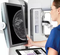
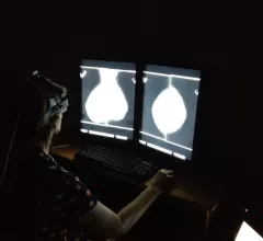
Breast imaging includes imaging modalities used for breast cancer screenings and planning therapy once cancer is detected. Mammography is the primary modality used. Mammogram technology is moving from 2D full-field digital mammography (FFDM) to breast tomosynthesis, or 3D mammography, which helps reduce false positive exams by allowing radiologists to look through the layers of tissue. Overlapping areas of dense breast tissue on 2D mammograms appear similar to cancers and 3D tomo helps determine if suspect areas are cancer or not. About 50% of women have dense breast tissue, which appears white on mammograms, the same as cancers, making diagnosis difficult. Radiologists use the Breast Imaging Reporting and Data System (BI-RADS) scoring system to define the density of breast tissue. Many states now require patients to be notified if they have dense breasts so they understand their mammograms might be suboptimal and they should use supplemental imaging that can see through the dense areas. This includes tomosythesis, breast ultrasound, automated breast ultrasound (ABUS), breast MRI, contrast enhanced mammography and nuclear imaging, including positron emission mammography (PEM).

healthcare value value-based care money dollar

Contrast-enhanced mammography for breast cancer staging offers significant cost savings

Prior research has shown that not only is contrast-enhanced spectral mammography comparable to CE-MRI in accuracy of loco-regional staging, but some studies have even found it to perform better.

Telemammography

4 key trends in breast imaging

These trends include growth in 3D mammography, supplemental imaging for women with dense breasts and in the role of artificial intelligence.

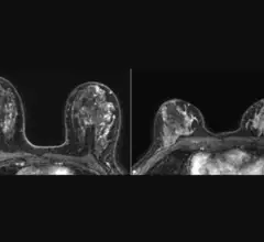
Example of various breast MRI protocol sequences that offer different types of soft tissue enhancement. Imaging performed on a Siemens Magnetom system. Breast MRI can help see through dense breast tissue to better detect cancers. #densebreasts #Breastdensity #BreastMRI

MRI a 'far superior' modality for dense breast imaging

The modality outperformed digital breast tomosynthesis, handheld ultrasound and automated breast ultrasound for cancer detection in a large cohort of women with dense breast tissue.

Thumbnail

Age and workload affect reader performance for screening mammograms

Age, specialty training and reading volume give readers an edge over their peers when it comes to screening mammogram interpretations.

Contrast-enhanced mammography uses iodine contrast injected into a patient and mammography system to image contrast uptake or areas of increased vascular activity, which is typical of cancers. This can help image through dense breast tissue to find cancers that are otherwise masked by dense breast tissue.

Radiologists should be aware of these clinical factors when reading CEM scans

The degree of background parenchymal enhancement on CEM during certain days of a premenopausal woman's cycle could impact how radiologists interpret the exam.

DBT guidance produces a significant drop in procedure time during radioseed localization

The savings in time and radiation dose do not come at the expense of surgical outcomes or complications.

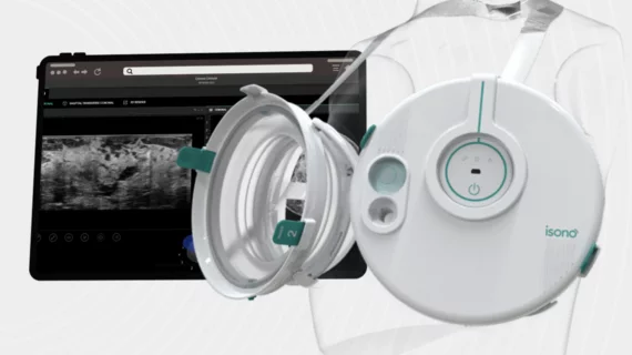
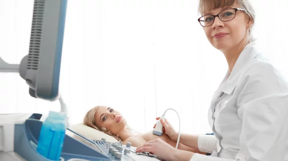
Automated breast ultrasound (ABUS) allows for reproducible breast imaging without variation based on which sonographer performs the exam. It also can help centers were they are short on qualified breast sonographers.  Breast ultrasound can help identify cancers, or benign cysts, even in women with very dense breast tissue. At the GE Healthcare booth at RSNA.

Commercially available AI systems excel in cancer detection in dense breasts

A multi-modal AI approach can combine information from both ABUS and DM, which could be especially beneficial in resource poor regions where experienced radiologists might not be readily available.

Example of artificial intelligence generated measurements to quantify the size of a lung cancer nodule during a followup CT scan to see if the lesion is regressing with treatment. This type of automation can aid radiologists by doing the tedious, time consuming work. Photo by Dave Fornell

8 trends in radiology technology to watch in 2023

Here is a list of some key trends in radiology technology from our editors based on our coverage of the radiology market.