Breast Imaging

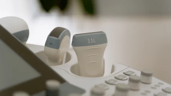
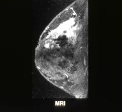
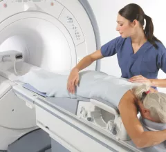
Breast imaging includes imaging modalities used for breast cancer screenings and planning therapy once cancer is detected. Mammography is the primary modality used. Mammogram technology is moving from 2D full-field digital mammography (FFDM) to breast tomosynthesis, or 3D mammography, which helps reduce false positive exams by allowing radiologists to look through the layers of tissue. Overlapping areas of dense breast tissue on 2D mammograms appear similar to cancers and 3D tomo helps determine if suspect areas are cancer or not. About 50% of women have dense breast tissue, which appears white on mammograms, the same as cancers, making diagnosis difficult. Radiologists use the Breast Imaging Reporting and Data System (BI-RADS) scoring system to define the density of breast tissue. Many states now require patients to be notified if they have dense breasts so they understand their mammograms might be suboptimal and they should use supplemental imaging that can see through the dense areas. This includes tomosythesis, breast ultrasound, automated breast ultrasound (ABUS), breast MRI, contrast enhanced mammography and nuclear imaging, including positron emission mammography (PEM).

Thumbnail

Breast MRI may help personalize treatment for DCIS patients

Pairing breast MRI with a test that characterizes breast cancer genes can lead to a more personalized treatment approach for patients with ductal carcinoma in situ (DCIS), reported authors of a recent study published in JAMA Oncology.

Thumbnail

Society of Breast Imaging addresses importance of screening among minority populations

The Society of Breast Imaging (SBI) addressed the importance of diversity in breast imaging in a recent statement published online Jan. 9, stating that breast cancer does not affect all ethnic and socioeconomic populations equally.

Thumbnail

SBI publishes policy statement on diversity, inclusion

The Society of Breast Imaging (SBI) has issued a new policy statement on diversity and inclusion, emphasizing the belief that “all people for whom breast cancer screening is appropriate should receive the opportunity to undergo screening.”

Thumbnail

Insurer's price calculator severely underestimates breast MRI cost for Philadelphia woman

Although many insurance providers have invested in price calculators for patients to determine out-of-pocket costs, one Philadelphia woman discovered they may severely underestimate actual prices for services, according to a recent report by The Philadelphia Inquirer.

Thumbnail

SBI, Oxford University Press to publish new Journal of Breast Imaging

On Wednesday, Oct. 10, the Society of Breast Imaging (SBI) and Oxford University Press (OUP) announced a partnership to publish the Journal of Breast Imaging (JBI)—the first peer-reviewed journal dedicated to breast imaging.

Thumbnail

Can diffusion-weighted imaging supplement dynamic contrast-enhanced breast MRI?

Diffusion-weighted imaging (DWI) could help differentiate between benign and malignant tumors and predict tumor recurrence in breast cancer patients, according to research published Oct. 4 in Academic Radiology. The authors noted that DWI may also serve as a beneficial supplement to contrast-enhanced breast MRI.

Thumbnail

Cardiac monitoring may protect high-risk breast cancer patients against heart failure

While heart failure (HF) is an uncommon complication of breast cancer, a new study in the Journal of the American College of Cardiology notes individuals treated with trastuzumab-based chemotherapy have a higher risk for HF—but they are not monitored for it, despite recommendations.

Thumbnail

Continued variation in radiology tech reports poses threat to readability

The lack of a structured reporting system for imaging technologists could be threatening the readability of studies in the field, a team of U.K. researchers reported this month in Radiography. But some argue a more rigid format would distract from the cognitive processes that make radiology reports so valuable in the first place.