Breast Imaging

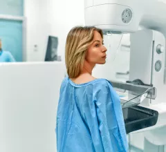
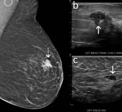
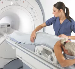
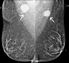
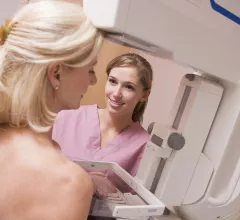
Breast imaging includes imaging modalities used for breast cancer screenings and planning therapy once cancer is detected. Mammography is the primary modality used. Mammogram technology is moving from 2D full-field digital mammography (FFDM) to breast tomosynthesis, or 3D mammography, which helps reduce false positive exams by allowing radiologists to look through the layers of tissue. Overlapping areas of dense breast tissue on 2D mammograms appear similar to cancers and 3D tomo helps determine if suspect areas are cancer or not. About 50% of women have dense breast tissue, which appears white on mammograms, the same as cancers, making diagnosis difficult. Radiologists use the Breast Imaging Reporting and Data System (BI-RADS) scoring system to define the density of breast tissue. Many states now require patients to be notified if they have dense breasts so they understand their mammograms might be suboptimal and they should use supplemental imaging that can see through the dense areas. This includes tomosythesis, breast ultrasound, automated breast ultrasound (ABUS), breast MRI, contrast enhanced mammography and nuclear imaging, including positron emission mammography (PEM).

Thumbnail

Providers are requesting fewer preoperative MRIs for breast cancer patients

Female surgeons, in particular, are abiding by evidence-based guidelines, but a more selective approach is still needed among doctors.

Thumbnail

Male breast cancer patients represent small portion of referrals but generate significant workload

About 75% of referrals result in an imaging exam, while only 1% lead to a cancer diagnosis, experts reported in Clinical Radiology.  

breast cancer mammography mammogram

‘Wake-up call’: Providers log substantially fewer breast biopsies with cancer diagnoses

The declines were more pronounced among Asian, Hispanic and Black women, imaging experts wrote in Radiology. 

5 Years into the Cloud, John Muir Health Is Just Getting Started

Sponsored by Sectra

One 3D mammogram acquired via digital breast tomosynthesis adds about 500 MB of image data to a hospital’s storage system. That’s the average. On the high end, a single study can occupy as much as 3 GB of real estate on a finite-volume storage server.

Chirag Pargh

Private equity-backed breast imaging giant names new CMO after leader departs for AI firm

Radiologist Chirag Parghi, MD, slid into the role in May following stints as interim CMO and director of breast imaging across several Houston-area locations. 

Thumbnail

Personalized breast cancer risk assessments at primary care up patients’ odds of mammography use

Annual mammography rates among women at high risk were significantly higher after the intervention, according to a new JAMA Network Open analysis. 

breast cancer mammography mammogram

Radiologists pilot immediate mammography reading, same-day recalls to boost breast cancer outcomes

Mass General physicians believe this practice is sustainable, resulting in less time spent on scheduling, reduced room cleaning, and lowering the chance of errors.

Thumbnail

Ditching paper for e-worklist translates to ‘substantially’ shorter wait times for breast imaging patients

Brigham and Women's recently started using an electronic health record-embedded tool, resulting in many improvements for preoperative breast localization procedures.