Breast Imaging

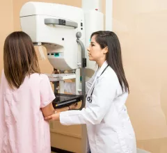
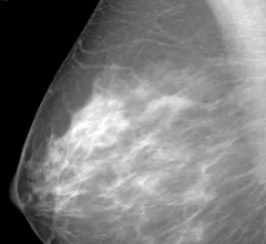
Breast imaging includes imaging modalities used for breast cancer screenings and planning therapy once cancer is detected. Mammography is the primary modality used. Mammogram technology is moving from 2D full-field digital mammography (FFDM) to breast tomosynthesis, or 3D mammography, which helps reduce false positive exams by allowing radiologists to look through the layers of tissue. Overlapping areas of dense breast tissue on 2D mammograms appear similar to cancers and 3D tomo helps determine if suspect areas are cancer or not. About 50% of women have dense breast tissue, which appears white on mammograms, the same as cancers, making diagnosis difficult. Radiologists use the Breast Imaging Reporting and Data System (BI-RADS) scoring system to define the density of breast tissue. Many states now require patients to be notified if they have dense breasts so they understand their mammograms might be suboptimal and they should use supplemental imaging that can see through the dense areas. This includes tomosythesis, breast ultrasound, automated breast ultrasound (ABUS), breast MRI, contrast enhanced mammography and nuclear imaging, including positron emission mammography (PEM).


Thumbnail

Machine learning model accurately predicts DCIS upstaging without invasive surgery

Understanding a patient's risk of developing invasive cancer without having to undergo surgery could help patients and providers choose more appropriate treatment plans.

breast ultrasound biopsy

Ultrasound outperforms four other modalities at assessing margins during breast surgery

Although ultrasound came out on top, achieving optimal operator performance could be taxing on resources, doctors cautioned.

breast radiologist breast cancer mammography

Key factors that influence radiology trainees’ interest in breast imaging

Repetitiveness is one of the most common reasons why residents and students avoid the subspecialty, according to new survey data.

Thumbnail

Deep learning rivals fellowship-trained radiologists at segmenting breast cancers on MRI

Researchers trained their platforms on more than 60,000 individual breast scans, significantly more than most architectures.

Thumbnail

Machine learning uses MRI to predict lymphovascular invasion in breast cancer patients

These algorithms could fill in where postoperative biopsy sometimes falls short, experts explained in Academic Radiology.

LGBTQ care lesbian gay rainbow

70% of breast radiologists surveyed either unsure about or had zero LGBTQ competency training

The finding is part of a survey of 400 breast imaging experts, published in the Journal of the American College of Radiology

breast cancer mammography mammogram

Survey explores radiology practices’ surveillance preferences when monitoring breast cancer survivors

There is “immense variability” in how this is handled in clinical practice, with a lack of evidence-based literature, experts wrote in JACR

Thumbnail

While promising, machine learning still misses 20% of cancers on breast MRIs, analysis shows

AI proved useful for detecting axillary lymph node metastases but isn’t yet ready for clinics, experts said recently.