X-ray

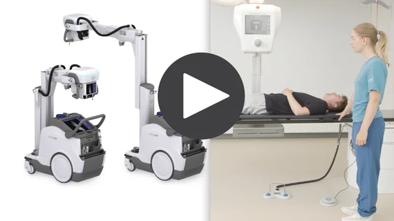
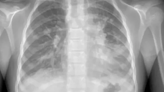
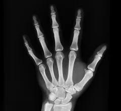
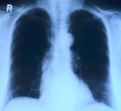
Medical X-rays have mostly converted from film, to computed radiography (CR) that used individual plates to record each X-ray digitally that then had to be uploaded into a PACS, to what is the standard-of-care today with digital radiography (DR). DR X-ray which allows immediate digital transfer of images into a PACS for immediate review. X-rays are used to diagnose fractures, bone abnormalities, lung pathologies and tumors, as well as monitor pediatric growth, plan for surgery and treat oncology patients during radiation therapy. More detailed anatomical imaging, especially soft tissue imaging, is usually sent for advanced imaging with CT or MRI. X-ray, especially mobile DR systems, are a primary use case for artificial intelligence (AI) integration. 

On review, popular imaging decision aid earns 1 thumbs-up—with caveats

With 91% sensitivity but only 25% specificity, the tool is worthwhile for clinicians who remain wary of frequent false positives that would send patients with no fractures for unneeded imaging.

Self-supervised AI ‘reads’ radiology reports to speed algorithm development

A machine learning system has come along that needs no human labeling of data for training yet matches radiologists at classifying diseases on chest X-rays—including some that the model was not specifically taught to detect.

Improving the diagnostic value of portable imaging: Experts propose 3 interventions

After experts from one institution evaluated 500 portable chest x-rays completed during the summer of 2021, it was revealed that 46.2% of the images obtained were problematic, requiring the imaging to be repeated. 

FDA clears X-ray system that produces hi-rez cine loops for chiropractors

Konica Minolta Healthcare has received FDA’s go-ahead to market a dynamic digital radiography (DDR) system.  

Thumbnail

TB-detecting AI tool shows promise for improving screening in low resource areas

A new artificial intelligence system can detect active tuberculosis on chest radiographs with accuracy comparable to radiologists, a recent paper in Radiology reports. 

Imaging fraud lands surgeon in jail

An orthopedic surgeon in California will spend seven years in prison for needlessly X-raying 10 patients hundreds of times over a four-year stretch.

The rise and fall of race-based radiation dosing: 4 lessons

Thanks to public outcry and legislative action in 1968, Black patients have not routinely received higher-dose X-rays than their fair-skinned peers for more than half a century.

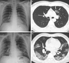
An overview of artificial intelligence (AI) in radiology with Keith Dreyer with the ACR. Images shows a COVID-19 lung CT scan reconstruction from Siemens Healthineers. #AI #radAI #ACR

AI speeds, improves chest X-ray interpretations

Six radiologists interpreting around 500 chest radiographs with an assist from AI bested unaided radiologists in measures of efficiency and/or accuracy in a new comparative performance study.