Magnetic Resonance Imaging

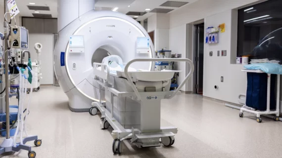
Magnetic resonance imaging (MRI) is the gold standard imaging modality for soft tissues. It produces detail cross-sectional images of soft tissue and bone anatomy, including muscles, tendons, ligaments, brain and organs, without the use of ionizing radiation. In addition to orthopedic imaging, MRI is also used for heart, brain and breast. MRI uses gadolinium contrast in many exams to highlight tissues and blood vessels, which enhances images and offers better diagnostic quality. It can also be used in conjunction with PET scans. How does MRI work? MR creates images by using powerful magnets to polarize hydrogen atoms in water (the body is made of of more than 80% water) so they face in one direction. A radiofrequency pulse is then used to ping these atoms, causing them to wobble, or resonate. The MRI coils detect this and computers can assemble images from the signals. Basic MRI scans will focus on the resonance of fat and water in two different sequences, which highlight and contrast different features in the anatomy.


MRI of knee with Osgood-Schlatter Disease

MRI scoring system simplifies diagnosis of common adolescent ortho issue

The condition can worsen with physical activity, which is why an accurate and timely diagnosis is important for healthy growth. 

birth control the pill cardiovascular disease blood clots side effects

Researcher scans herself 75 times to study effects of birth control on brain health

The postdoctoral research fellow hopes her research will provide greater insight into how the use of oral birth control impacts physical and mental health.

Thumbnail

Brain changes related to Huntington's evident on imaging decades before symptoms emerge

Experts hope their findings offer valuable insights that one day lead to improved interventions aimed at preserving brain function in patients with Huntington's. 

Thumbnail

New GBCA uses 60% less gadolinium

Bayer’s latest contrast agent is proving its potential in clinical trials as a promising alternative to agents with higher gadolinium content. 

Thumbnail

Advanced MRI techniques may provide insight into brain damage stemming from youth sports

The study is part of a bigger effort to understand why some children recover better than others following head injuries.

Thumbnail

Basketball players' brains are wired differently, new MRI data suggest

Although some form of neural superiority may not be the driving factor behind an elite athlete’s talent, new evidence shows these individuals have unique structure and connectivity patterns.

Thumbnail

Altered brain structure may play a role in eating disorders, MRI data suggest

Experts recently uncovered evidence suggesting  behaviors such as undereating, binging and purging could be owed to anatomical alterations in parts of the brain. 

prostate cancer PSA

Payment incentives fail to push physicians toward MRI surveillance of low-risk prostate cancer

BCBS of Michigan has sought to push urologists away from aggressive treatment for low-risk cancer, offering a payment perk to shift their approach.