Magnetic Resonance Imaging

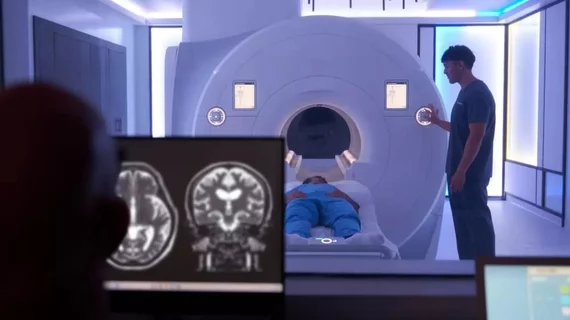
Magnetic resonance imaging (MRI) is the gold standard imaging modality for soft tissues. It produces detail cross-sectional images of soft tissue and bone anatomy, including muscles, tendons, ligaments, brain and organs, without the use of ionizing radiation. In addition to orthopedic imaging, MRI is also used for heart, brain and breast. MRI uses gadolinium contrast in many exams to highlight tissues and blood vessels, which enhances images and offers better diagnostic quality. It can also be used in conjunction with PET scans. How does MRI work? MR creates images by using powerful magnets to polarize hydrogen atoms in water (the body is made of of more than 80% water) so they face in one direction. A radiofrequency pulse is then used to ping these atoms, causing them to wobble, or resonate. The MRI coils detect this and computers can assemble images from the signals. Basic MRI scans will focus on the resonance of fat and water in two different sequences, which highlight and contrast different features in the anatomy.

Thumbnail

GBCA dose reduced by up to 80% with help from deep learning-based image reconstruction

Although GBCAs are largely considered safe, there are concerns about how gadolinium retention could affect patients who require repeated imaging.

Siemens Healthineers

Siemens Healthineers inks 10-year, $105M imaging value partnership with large medical center

The deal is with the Ohio State Wexner Medical Center, covering equipment purchases and consulting services. 

Thumbnail

Vaping may affect vascular function as much or more than smoking, MRI study shows

E-cigarettes, or vapes, are largely considered a safer alternative to smoking tobacco cigarettes, but new MRI data suggest they still pose significant risk to users’ health. 

technologist MRI rad tech radiologic radiology

CMS approves new MRI standards related to remote scanning

Accrediting body IAC said revisions help imaging facilities ensure the safe delivery of MRIs, including when key personnel are located off-site.

technologist MRI rad tech radiologic radiology

Deep learning-based reconstruction nearly halves spinal MRI acquisition times

The accelerated exam does not come at the expense of image quality, according to new work published in Academic Radiology. 

doctor examines patient data on their tablet

Radiologists are more accurate, confident when they know the full story

Providing radiologists with key details about the patient's symptoms can go a long way toward improving the quality of their work. 

Thumbnail

MRI guidance significantly reduces radiotherapy side effects compared to CT

A recent comparison of the two modalities’ utility for guiding radiation therapy for prostate cancer shows numerous benefits for MRI over CT.

Thumbnail

Socioeconomic disadvantage during childhood linked to altered brain structure later in life, MRIs show

New MRI data highlights associations between being born into disadvantaged communities and decreased volume of multiple areas in the brain. What’s more, these findings hold true despite educational attainment and late-life income.