Magnetic Resonance Imaging

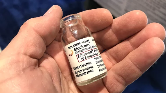
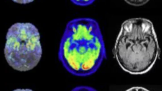
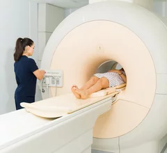
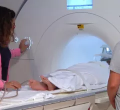
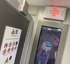
Magnetic resonance imaging (MRI) is the gold standard imaging modality for soft tissues. It produces detail cross-sectional images of soft tissue and bone anatomy, including muscles, tendons, ligaments, brain and organs, without the use of ionizing radiation. In addition to orthopedic imaging, MRI is also used for heart, brain and breast. MRI uses gadolinium contrast in many exams to highlight tissues and blood vessels, which enhances images and offers better diagnostic quality. It can also be used in conjunction with PET scans. How does MRI work? MR creates images by using powerful magnets to polarize hydrogen atoms in water (the body is made of of more than 80% water) so they face in one direction. A radiofrequency pulse is then used to ping these atoms, causing them to wobble, or resonate. The MRI coils detect this and computers can assemble images from the signals. Basic MRI scans will focus on the resonance of fat and water in two different sequences, which highlight and contrast different features in the anatomy.

Deep learning application spots acute ischemic stroke in less than 30 seconds on MRI

What’s more, the application’s performance was accurate, even when fewer scanning sequences were utilized.

Thumbnail

MRI findings predict Parkinson's disease progression

MR imaging could provide insight into how Parkinson’s disease will progress years before its related symptoms become debilitating.  

Murderer's fate hinges on 2 different interpretations of his brain scans

A radiologist’s testimony could deconstruct a legal team’s argument that their defendant had a brain tumor that may have affected his behavior when he shot and killed five women in 2019. 

Thumbnail

How having ovaries removed before menopause affects brain structure

These changes are especially prevalent in women who have the surgery before the age of 40, new research suggests. 

Thumbnail

Fluorinated contrast agents show 'strong potential' for MRI theranostics

Experts have developed a first-of-its-kind fluorinated contrast agent they believe will provide more detailed MR imaging and aid in the treatment of certain cancers. 

Thumbnail

Delays are common with sedated MRIs. Standardized workflow protocols could help avoid them

In some cases, MRI with sedation can delay workflows by 60 minutes or more, putting staff and patients in a bind.

How patients' focus affects data derived from functional MRI scans

As a person’s concentration dwindles during resting state or task-based sequences, the resultant brain activity observed on imaging could be misleading.

radiology match day fellowship

Following 2024 fellowship matching, neuroradiology said to be ‘at its hottest point ever’

Neuroradiology has topped the field among imaging subspecialties vying for new fellows to train, filling 287 of 316 certified positions—a 90.8% fill rate—across 87 fellowship programs.