Magnetic Resonance Imaging

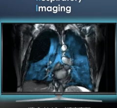
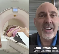
Magnetic resonance imaging (MRI) is the gold standard imaging modality for soft tissues. It produces detail cross-sectional images of soft tissue and bone anatomy, including muscles, tendons, ligaments, brain and organs, without the use of ionizing radiation. In addition to orthopedic imaging, MRI is also used for heart, brain and breast. MRI uses gadolinium contrast in many exams to highlight tissues and blood vessels, which enhances images and offers better diagnostic quality. It can also be used in conjunction with PET scans. How does MRI work? MR creates images by using powerful magnets to polarize hydrogen atoms in water (the body is made of of more than 80% water) so they face in one direction. A radiofrequency pulse is then used to ping these atoms, causing them to wobble, or resonate. The MRI coils detect this and computers can assemble images from the signals. Basic MRI scans will focus on the resonance of fat and water in two different sequences, which highlight and contrast different features in the anatomy.

Thumbnail

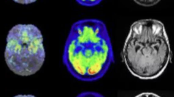
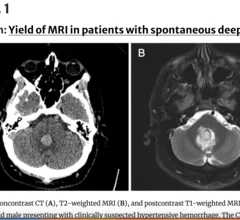
Even two years after recovery, damage to multiple regions of the brain persists in COVID patients

On imaging some former COVID patients show significant alterations in several parts of the brain related to emotional regulation.

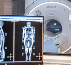
GE Health and MedStar Health are collaborating with the National Basketball Association (NBA), National Basketball Players Association (NBPA) and the Next Gen Basketball Players Union (NBGPU)

NBA partners with GE HealthCare, MedStar Health to gain imaging insights into elite athletes' health

The goal of the study is to obtain quantitative data on how daily training and competing affect players’ musculoskeletal health. The information will be used to inform training, rest and recovery periods. 

Siemens Healthineers

Siemens Healthineers investing $314M to build new MRI manufacturing facility

This is the first major production site for the company's DryCool technology, which drastically reduces the amount of helium required for magnetic resonance imaging. 

Thumbnail

New low-field MRI small enough to plug into a standard wall outlet saves money without sacrificing safety

And with the help of deep learning techniques, it provides diagnostic quality imaging.

prostate cancer PSA

Collaborative bolsters prostate MRI quality, with 1 site realizing $559,000 in reimbursement gains

Some of the most commonly deployed interventions included protocol adjustments, standardized patient prep instructions and personnel training. 

How beneficial is MR-directed breast ultrasound in reducing biopsies?

A team of experts determined that correlating masses initially detected on MRI are significantly more likely to result in a cancer diagnosis than other common findings. 

Thumbnail

Point-based risk prediction model reduces prostate biopsies by up to 20%

The model incorporates specific data from MRI exams with patient risk factors to predict whether a person is likely to develop clinically significant prostate cancer.

live MRI video of stuttering

Real-time MRI shows exact mechanisms underlying man's stutter

This development could help scientists fine-tune speech therapy to people who struggle with speaking.