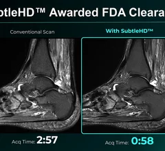
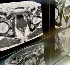
Magnetic Resonance Imaging

Magnetic resonance imaging (MRI) is the gold standard imaging modality for soft tissues. It produces detail cross-sectional images of soft tissue and bone anatomy, including muscles, tendons, ligaments, brain and organs, without the use of ionizing radiation. In addition to orthopedic imaging, MRI is also used for heart, brain and breast. MRI uses gadolinium contrast in many exams to highlight tissues and blood vessels, which enhances images and offers better diagnostic quality. It can also be used in conjunction with PET scans. How does MRI work? MR creates images by using powerful magnets to polarize hydrogen atoms in water (the body is made of of more than 80% water) so they face in one direction. A radiofrequency pulse is then used to ping these atoms, causing them to wobble, or resonate. The MRI coils detect this and computers can assemble images from the signals. Basic MRI scans will focus on the resonance of fat and water in two different sequences, which highlight and contrast different features in the anatomy.

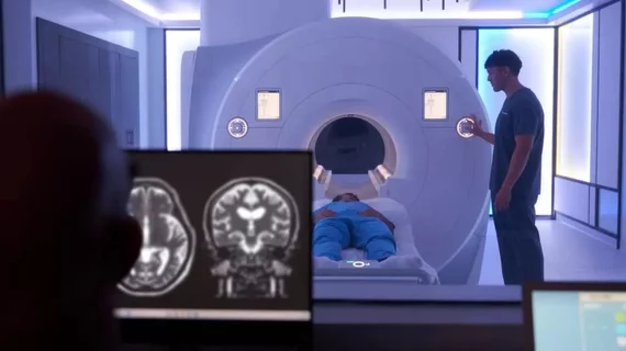
Auburn University SIemens 7T magnetom scanner

University charts a world’s first with clinical use of new $9M 7T MRI system

The Siemens Magnetom scanner utilizes dedicated radiofrequency sodium coils and parallel transmit technology—a “stark” difference from older models

Thumbnail

Deep learning-based MRI reconstruction software produces considerable cost savings

Oulu University Hospital believes AI will allow it to deliver the same level of service with one fewer scanner, enabling annual savings of over $436,000.

The rapid rise of artificial intelligence (AI) has helped cardiologists, radiologists, nurses and other healthcare providers embrace precision medicine in a way that ensures more heart patients are receiving personalized care.

FDA clears AI-enhanced 3D heart models generated from MR and CT images

The inHeart models significantly reduce cardiac ablation surgery times.

Thumbnail

NIH study finds no evidence of ‘Havana syndrome’ in brains of affected individuals

Brain scans on those with symptoms of the mysterious illness and a control group were found to be the same.

Radiologist using the Philips Smart Quant 3D Neuro artificial intelligence (AI) software to perform our measurements for white matter, gray matter and other parameters on brain MRI.

PHOTO GALLERY of brain imaging

This is a clinical photo gallery of neuro imaging and what conditions can be can be visualized in brain scans, and various imaging techniques used.

Medical 3D printing expert Summer Decker, PhD, professor and vice chair for research and innovation, and director of 3D clinical applications, University of South Florida (USF) Morsani College of Medicine and Tampa General Hospital, explains what is involved in creating a 3D printiung program in radiology and how it helps patient care.

Pitching the business case for 3D printing labs in radiology

Medical 3D printing expert Summer Decker, PhD, University of South Florida, explains how her lab has helped improve patient care. 

Prenuvo MRI Sollis Health

Whole-body MRI startup Prenuvo partners with concierge emergency care provider catering to the wealthy

Sollis Health operates private emergency rooms, allowing customers to bypass hospitals to receive around-the-clock care from physicians. 

The 5-minute MRI: AI algorithm reduces scan times by 57% while maintaining image quality

The decrease in duration allows for more examinations in the same timeframe, enhances patient comfort and reduces the chance of image distortion.