Magnetic Resonance Imaging

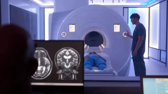
Magnetic resonance imaging (MRI) is the gold standard imaging modality for soft tissues. It produces detail cross-sectional images of soft tissue and bone anatomy, including muscles, tendons, ligaments, brain and organs, without the use of ionizing radiation. In addition to orthopedic imaging, MRI is also used for heart, brain and breast. MRI uses gadolinium contrast in many exams to highlight tissues and blood vessels, which enhances images and offers better diagnostic quality. It can also be used in conjunction with PET scans. How does MRI work? MR creates images by using powerful magnets to polarize hydrogen atoms in water (the body is made of of more than 80% water) so they face in one direction. A radiofrequency pulse is then used to ping these atoms, causing them to wobble, or resonate. The MRI coils detect this and computers can assemble images from the signals. Basic MRI scans will focus on the resonance of fat and water in two different sequences, which highlight and contrast different features in the anatomy.

An MRI of a fetus inside the mother. The freakish appearance of the eyes and the face are normal for MRI fetal imaging. Image courtesy of RSNA

PHOTO GALLERY: What does fetal medical imaging look like?

This is a clinical photo gallery of fetal imaging that explains what all can be seen on medical imaging, how sex is determined, how measurements are used to track the development of a baby.

Researchers develop new method for early detection of Alzheimer’s

Higher levels of amyloid protein in the blood correlate with MRI images of the brain that are consistent with dementia.

MRI anxiety worse for girls from minority backgrounds, study finds

Latina girls ages 8-13 were placed in an MRI and given an emotional processing test to measure fear response.

Kate Hanneman, MD, University Of Toronto, explains why vendors and hospitals are increasingly discussing lowing their carbon footprint by starting with radiology. 

What does radiology have to do with climate change?

Kate Hanneman, MD, explains why many vendors and hospitals want to lower radiology's impact on the environment. "Taking steps to reduce the carbon footprint in healthcare isn’t just an opportunity," she said. "It’s also a responsibility."

Thumbnail

SimonMed imaging centers avoided regulation, even after safety concerns

The chain of imaging centers was given a private practice exemption in Arizona, but it includes more than 60 different facilities.

ACR updates requirements for on-site staff overseeing contrast administration

A clinician trained to manage reactions should be on-site at any imaging facilities doing contrast studies to maintain patient safety.

cocaine heart failure HFrEF LVEF hospitalizations beta-blockers hypertension cardiology

MRI scans on rat brains could lead to new cocaine addiction therapies

A study on rat models reveals how prolonged cocaine use disrupts neural networks responsible for regulating emotions and problem solving.

Cardiologist heart

Cardiac imaging use is skyrocketing, and radiologists are rising to the challenge

Over the course of a decade, the number of CT exams leapt 353% by 2022, while MRI climbed nearly 283%, researchers detailed Wednesday.