Magnetic Resonance Imaging

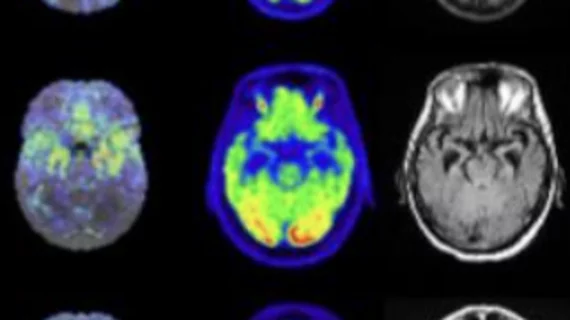
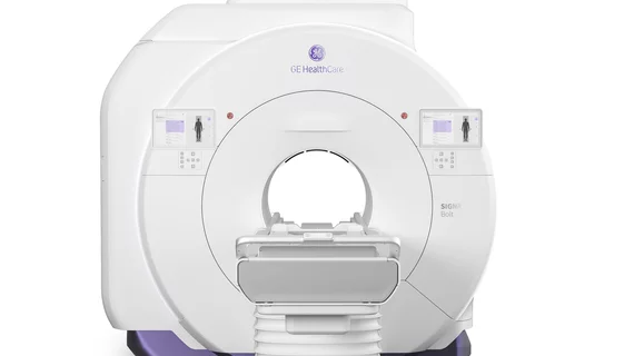
Magnetic resonance imaging (MRI) is the gold standard imaging modality for soft tissues. It produces detail cross-sectional images of soft tissue and bone anatomy, including muscles, tendons, ligaments, brain and organs, without the use of ionizing radiation. In addition to orthopedic imaging, MRI is also used for heart, brain and breast. MRI uses gadolinium contrast in many exams to highlight tissues and blood vessels, which enhances images and offers better diagnostic quality. It can also be used in conjunction with PET scans. How does MRI work? MR creates images by using powerful magnets to polarize hydrogen atoms in water (the body is made of of more than 80% water) so they face in one direction. A radiofrequency pulse is then used to ping these atoms, causing them to wobble, or resonate. The MRI coils detect this and computers can assemble images from the signals. Basic MRI scans will focus on the resonance of fat and water in two different sequences, which highlight and contrast different features in the anatomy.


Thumbnail

Dozens of Indigenous Canadians file lawsuit against two radiologists over undisclosed study

The plaintiffs claim they were imaged for a liver disease study without giving their consent, or learning of the results. 

Thumbnail

MRI reveals placental oxygenation levels may be linked to fetal brain development

Oxygenation levels not only seem to be a predictor of cortical growth and cognition, but likely childhood behavior as well, the study found. 

Thumbnail

Class action lawsuit claims radiologists subjected patients to ‘secret’ MRI research

Physician allegedly kept patients in scanners for extra examinations as part of a study evaluating the livers of indigenous peoples. 

stroke brain dementia alzheimer's puzzle mental health

MRI shows brain ‘leakage’ that may explain cognitive decline from long COVID

A team from Ireland identified disruptions to the integrity of the blood vessels in the brains of patients experiencing brain fog due to the disease.

Avatar Medical virtual reality imaging

Startup that creates avatars via CT, MR images raises $5.4M from radiologists and investors

Paris-based Avatar Medical believes such physician financing demonstrates “strong support” for its medical solution.

An example of cardiac MRI myocardial blood flow reserve (MBFR) imaging. Image courtesy of https://doi.org/10.3390/diagnostics10090679

Imaging group thrilled with new CPT codes for MBFR measurements

The Society for Cardiovascular Magnetic Resonance applauded two new CPT Category III codes for myocardial blood flow reserve (MBFR) assessments.

Thumbnail

Microrobots guided by an MRI eradicate liver cancer

The technique was successful in a proof-of-concept animal study, but human trials are a long way off. 

Price shopping transparency

‘MRI’ is the most-searched medical term among patients seeking cost information

“CT scan” was the third most-searched term among healthcare consumers, and “ultrasound” was No. 4, according to Fair Health.