Magnetic Resonance Imaging

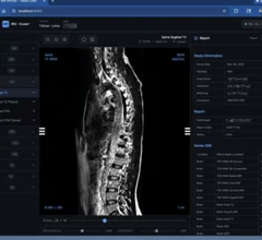
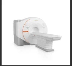
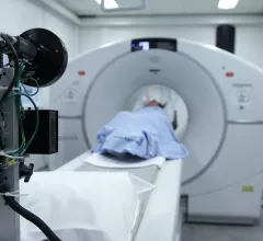
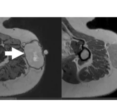
Magnetic resonance imaging (MRI) is the gold standard imaging modality for soft tissues. It produces detail cross-sectional images of soft tissue and bone anatomy, including muscles, tendons, ligaments, brain and organs, without the use of ionizing radiation. In addition to orthopedic imaging, MRI is also used for heart, brain and breast. MRI uses gadolinium contrast in many exams to highlight tissues and blood vessels, which enhances images and offers better diagnostic quality. It can also be used in conjunction with PET scans. How does MRI work? MR creates images by using powerful magnets to polarize hydrogen atoms in water (the body is made of of more than 80% water) so they face in one direction. A radiofrequency pulse is then used to ping these atoms, causing them to wobble, or resonate. The MRI coils detect this and computers can assemble images from the signals. Basic MRI scans will focus on the resonance of fat and water in two different sequences, which highlight and contrast different features in the anatomy.

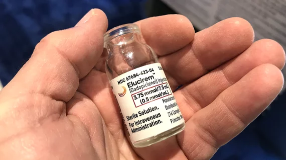
Could China's latest restrictions impact the supply of MRI contrast?

The ongoing trade war between the United States and China could jeopardize the availability of contrast agents used for medical imaging. 

Imricor Medical Systems ventricular ablation

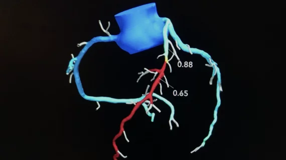
Cardiologists make history, perform world’s first ventricular ablation guided by real-time MRI

“MRI images provide a better view of the anatomy of the heart and the instruments used for treatment, but also of the changes made to the treated cardiac tissue,” one cardiologist explained. “Unlike X-ray images, the entire area surrounding the heart can be seen, including the blood vessels and valves."

Thumbnail

Reducing MRI scan times could add thousands of exam slots, generating up to $22M in new revenue per practice

That’s according to a new retrospective study led by the University of California, San Francisco, and Siemens Healthineers, published Tuesday in Radiology

Thumbnail

Imaging shows why music makes us happy

Experts believe their findings could be significant for treating both pain and mental health disorders. 

scanner money radiology dollar MRI CT payment compensation

MRI projectile incident likely to result in a 'seven-figure sum' repair bill for hospital

News of an incident is a stark reminder that healthcare workers and patients aren’t the only ones who need to be aware around MRI suites.

Thumbnail

Experts may have discovered why some patients are more susceptible to GBCA-related side effects

A new study focuses on the role oxalic acid may play in the body’s ability to eliminate gadolinium after an MRI study.

istock-1359148358.jpg

Radiology groups spar over certificate-of-need application for $1.7M new MRI scanner

Diagnostic Imaging of Southbury recently asked for permission to buy a second MR system, with its first currently operating at 120% capacity. 

Multiple sclerosis ribbon MS

AI tool has 'untapped potential' for diagnosing and managing multiple sclerosis

The tool extracts brain region and white matter lesion volumes from any MRI of the brain, even less specialized sequences not traditionally used in the diagnosis or management of MS.