Magnetic Resonance Imaging

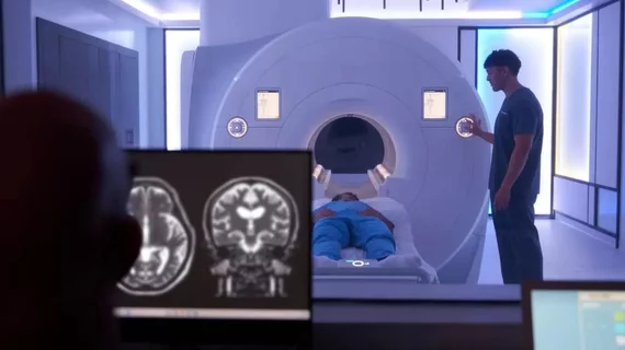
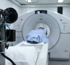
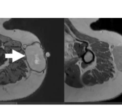
Magnetic resonance imaging (MRI) is the gold standard imaging modality for soft tissues. It produces detail cross-sectional images of soft tissue and bone anatomy, including muscles, tendons, ligaments, brain and organs, without the use of ionizing radiation. In addition to orthopedic imaging, MRI is also used for heart, brain and breast. MRI uses gadolinium contrast in many exams to highlight tissues and blood vessels, which enhances images and offers better diagnostic quality. It can also be used in conjunction with PET scans. How does MRI work? MR creates images by using powerful magnets to polarize hydrogen atoms in water (the body is made of of more than 80% water) so they face in one direction. A radiofrequency pulse is then used to ping these atoms, causing them to wobble, or resonate. The MRI coils detect this and computers can assemble images from the signals. Basic MRI scans will focus on the resonance of fat and water in two different sequences, which highlight and contrast different features in the anatomy.

Aclarion, a company using remote MRIs to assess back pain, expands in partnership with RadNet

The publicly traded Broomfield, Colorado, firm plans to bolster access to its Nociscan product across key markets in New York and New Jersey. 

Experts identify 'critical window' when early signs of neurodegeneration become visible on imaging

These findings may lead to new screening guidelines that could change how imaging is utilized in the aging population.

UM Health Care's 7T MRI scanner receives ACR accreditation

University of Missouri Health Care's 7T scanner joins exclusive ACR accreditation club

There are fewer than 40 of these 7T MRI scanners in North America, and MU Health Care’s is one of less than 10 to achieve ACR accreditation.

VI-RADS threshold, imaging features predict bladder cancer invasiveness with nearly 100% accuracy

New findings related to Vesical Imaging-Reporting and Data System scores and specific MRI findings could improve the management of bladder cancer. 

FCI scanner more ably detects cancer spread than traditional MRI

New low-field scanner detects cancer spread better than traditional breast MRI

Researchers involved in its development are hopeful that the scanner could eventually lead to improved outcomes in cancer patients who require surgery to remove malignant tissue. 

David A. Rosman, MD, MBA, deputy chief, radiology enterprise service, Mass General Brigham, explains details of a recent AJR article that showed imaging outside of hospitals could potentially save billions.

Shift toward imaging outside the hospital could save billions

Harvard’s David A. Rosman, MD, MBA, explains how moving imaging outside of hospitals could save billions of dollars for U.S. healthcare.

PHOTO GALLERY: thyroid nodule imaging

This is a collection of radiology imaging showing various presentations of thyroid nodules.

Biograph whole-body MRI

Biograph, a new whole-body MRI startup, launches with $7,500 membership fee

The company has already opened its first location in San Mateo, California, with another to follow in New York City, collecting over 1,000 datapoints from 30-plus exams.