X-ray

Medical X-rays have mostly converted from film, to computed radiography (CR) that used individual plates to record each X-ray digitally that then had to be uploaded into a PACS, to what is the standard-of-care today with digital radiography (DR). DR X-ray which allows immediate digital transfer of images into a PACS for immediate review. X-rays are used to diagnose fractures, bone abnormalities, lung pathologies and tumors, as well as monitor pediatric growth, plan for surgery and treat oncology patients during radiation therapy. More detailed anatomical imaging, especially soft tissue imaging, is usually sent for advanced imaging with CT or MRI. X-ray, especially mobile DR systems, are a primary use case for artificial intelligence (AI) integration. 

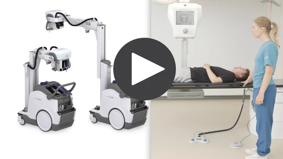
The Nanox ARC cold-cathode, tomosythesis 3D X-ray system is currently pending FDA clearance. If cleared, it would be the first device of its kind to offer cold-cathode X-ray tube technology and the ability to slice through the anatomy in images similar to CT scans aid diagnosis. The new type of tube also could greatly reduce the size and weight of X-ray systems. The vendor sees this system as key to its larger plan to address health disparities and access to imaging world-wide. #Nanox

Can cold-cathode X-ray combined with teleradiology and AI eliminate health disparities?

The Israeli vendor Nanox says it has a vision for the future of healthcare. It seeks to address health disparities and access challenges with a new business model and innovative package of technologies. Hurdles loom, but opportunities abound. 

Thumbnail

Dynamic chest radiography a suitable, low-cost alternative to V/Q scanning for pulmonary hypertension

Dynamic chest radiography was recently shown to be comparable to lung ventilation-perfusion scanning for detecting chronic thromboembolic pulmonary hypertension.  

hand x-ray radiograph

Has electronic cropping in digital radiography resulted in the death of collimation?

While electronically cropping an image may seem like a harmless act, the habit is not without unintended consequences, the authors of a new paper recently explained.

Thumbnail

Chest X-rays alone ineffective in COVID triage

New research indicates that there is significant reader variability in COVID classifications among different specialties when chest X-rays alone are the diagnostic tool of choice.

X-rays may suffice for assessing some low-back fractures

It’s unlikely radiography will unseat MRI for routinely distinguishing between acute and chronic compression fractures of the lumbar vertebrae, but certain X-ray findings might, in cases, obviate the need for the pricier imaging option.

‘Radiology failures, misdiagnosed fractures’ blamed in 2 wrongful assumptions of child battery

In the U.K., two instances of evidently inept work by radiologists are inadvertently spotlighting the value of subspecialized image interpretation in socially sensitive patient cases.

Mr. O’Lear receives his sentence: 15 years and $2M

The criminal saga of the X-ray business owner who got caught fleecing CMS of $2 million—while grabbing for another $1.7 million or so—has reached its epilogue.

Representative cases showing pneumonia extents and patterns on chest X-ray (CXRs) and CT images. (E and F) A 36-year-old male with no history of vaccination for COVID- 19. The patient had no history of comorbidity. Axial chest CT image obtained on the same day showing unilateral ground-glass opacity with a nonrounded morphology and non-peripheral distribution in the left upper lobe (arrows). RSNA Image. COVID on X-ray, CT scan. What does COVID look like in medical imaging? Example of COVID imaging.

Vaccinated vs unvaccinated, Delta vs Omicron—how do these factors impact clinical and imaging features?

Vaccination rates likely had a role in reducing disease severity during Omicron, a new paper published in Radiology suggests.