Computed Tomography

Computed tomography (CT) is a fast and accurate imaging modality often used in emergency settings and trauma imaging. CT scans, with or without (or both) iodinated contrast are frequently used to image the brain, chest, abdomen and pelvis, but also have post-imaging reconstructive capabilities for detailed orthopedic imaging. It is now a standard imaging modality in emergency rooms to quickly assess patients. CT uses a series of X-ray images shot as the gantry rotates around the patient. Computer technology assembles these into into a dataset volume than can be slices on any access, or advanced visualization software can extract specific parts of the anatomy for study. Find more content specific to cardiac CT.

Thumbnail

Incidental finding characteristics that warrant communication

Three factors are associated with true high-risk findings—imaging protocols for aortic valve implantation planning, imaging area, and cardiology department orders.

Left, coronary CT angiography of a vessel showing plaque heavy calcium burden. Right, image showing color code of various types of plaque morphology showing the complexity of these lesions. The right image was processed using the FDA cleared, AI-enabled plaque assessment from Elucid.

FDA clears new software for AI-powered CCTA assessments

Elucid's PlaqueIQ was trained to turn CCTA images into interactive 3D reports that help physicians visualize the presence of atherosclerosis.

Deep learning denoising produces quality trauma head CT images at 25% of the radiation dose

Denoising algorithm produces quality trauma head CTs at a fraction of the radiation dose

Radiation doses needed for quality head CT scans in trauma settings can be reduced by as much as 75% using DL-based denoising.

CTC colonography colon cancer colonoscopy

ACR concerned pay cap on CT-based colon cancer screening could make service ‘untenable’

Medicare wants to limit the technical component for the exam to the HOPPS rate, which would represent an 81% reduction to the MPFS amount of $459.92. 

The rapid rise of artificial intelligence (AI) has helped cardiologists, radiologists, nurses and other healthcare providers embrace precision medicine in a way that ensures more heart patients are receiving personalized care.

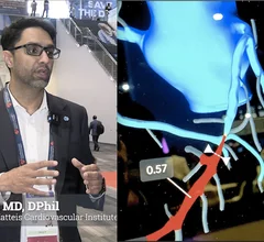
FDA clears AI platform for planning ahead of TAVR, other heart procedures

The cloud-based platform identifies and measures cardiac structures in CT scans.

SCCT 2024-2025 President Maros Ferencik (right) shared an office with SCCT's first president Stephan Achenbach (left) in 2002.

New SCCT president takes office, lists priorities

Maros Ferencik, MD, noted that the Society of Cardiovascular Computed Tomography represents specialists from many different specialties, including cardiology, radiology and beyond. 

CT scans highlight impact of semaglutide on body composition

Body composition metrics derived from CT scans provide greater insight into how losing weight affects the body from a cardiometabolic perspective.

Thumbnail

Guided errors in medical training improve interpretation skills post-residency

A new study in JAMA delves into how error management training builds adaptive expertise in interpreting head CT studies among nonradiologists.