Computed Tomography

Computed tomography (CT) is a fast and accurate imaging modality often used in emergency settings and trauma imaging. CT scans, with or without (or both) iodinated contrast are frequently used to image the brain, chest, abdomen and pelvis, but also have post-imaging reconstructive capabilities for detailed orthopedic imaging. It is now a standard imaging modality in emergency rooms to quickly assess patients. CT uses a series of X-ray images shot as the gantry rotates around the patient. Computer technology assembles these into into a dataset volume than can be slices on any access, or advanced visualization software can extract specific parts of the anatomy for study. Find more content specific to cardiac CT.

Radiologist using the Philips Smart Quant 3D Neuro artificial intelligence (AI) software to perform our measurements for white matter, gray matter and other parameters on brain MRI.

PHOTO GALLERY of brain imaging

This is a clinical photo gallery of neuro imaging and what conditions can be can be visualized in brain scans, and various imaging techniques used.

March 18, 2024

Outpatient study finds CT contrast is generally safe, but risk of adverse events varies with the seasons

A three-institution study of 473,482 patients found the risk of an adverse event following CT contrast increases in the summer and fall.

March 15, 2024
quality imaging appropriateness clinical decision support CAS AUC

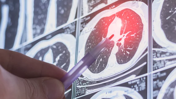
Key factors help AI predict whether patients will follow up for incidental lung nodules

A new JACR study examines 13 potential predictors and their influence on patients' actions after their CT scan unearthed unexpected findings. 

March 15, 2024
Medical 3D printing expert Summer Decker, PhD, professor and vice chair for research and innovation, and director of 3D clinical applications, University of South Florida (USF) Morsani College of Medicine and Tampa General Hospital, explains what is involved in creating a 3D printiung program in radiology and how it helps patient care.

Pitching the business case for 3D printing labs in radiology

Medical 3D printing expert Summer Decker, PhD, University of South Florida, explains how her lab has helped improve patient care. 

March 14, 2024

New imaging protocols proposed to curb rise of cardiovascular infections

Eleven medical societies have signed on to a consensus statement aimed at standardizing imaging for suspected cardiovascular infections.

March 12, 2024
An MRI of a fetus inside the mother. The freakish appearance of the eyes and the face are normal for MRI fetal imaging. Image courtesy of RSNA. Baby MRI, baby ultrasound

PHOTO GALLERY: What does fetal medical imaging look like?

This is a clinical photo gallery of fetal imaging that explains what all can be seen on medical imaging, how sex is determined, how measurements are used to track the development of a baby.

March 11, 2024
Fish

Museum project uses contrast CT and X-ray to catalog world’s vertebrates

Over the course of five years, 18 different institutions worked to produce the images for a Florida Museum initiative.

March 6, 2024
Using computed tomography (CT) to perform coronary artery calcium (CAC) scoring can help identify symptomatic chest pain patients who do not require further testing, according to a new analysis published in Radiology.[1]

Coronary calcium scoring predicts when chest pain patients can skip invasive testing

Using CT to perform coronary artery calcium scoring on symptomatic chest pain patients can deliver significant value, according to a new data published in Radiology

March 6, 2024