Computed Tomography

Computed tomography (CT) is a fast and accurate imaging modality often used in emergency settings and trauma imaging. CT scans, with or without (or both) iodinated contrast are frequently used to image the brain, chest, abdomen and pelvis, but also have post-imaging reconstructive capabilities for detailed orthopedic imaging. It is now a standard imaging modality in emergency rooms to quickly assess patients. CT uses a series of X-ray images shot as the gantry rotates around the patient. Computer technology assembles these into into a dataset volume than can be slices on any access, or advanced visualization software can extract specific parts of the anatomy for study. Find more content specific to cardiac CT.

Thumbnail

How radiologists can help identify victims of domestic violence

New analysis details patterns radiologists can look for that may signal a patient is experiencing domestic violence. 

Thumbnail

Using CT imaging to opportunistically screen for osteoporosis could save Medicare upward of $2.5B

Only about 11% to 18% of eligible patients receive recommended bone density scans, but CT images gathered for other purposes could fill this gap. 

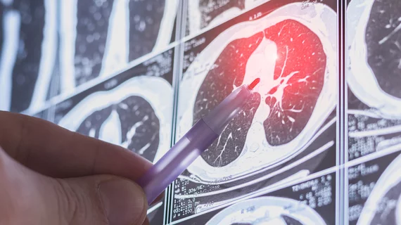
AI reduces CT lung cancer screening workload by nearly 80%

And it can do so with almost 100% accuracy as a first reader, according to a new large-scale analysis.

David A. Rosman, MD, MBA, deputy chief, radiology enterprise service, Mass General Brigham, explains details of a recent AJR article that showed imaging outside of hospitals could potentially save billions.

Shift toward imaging outside the hospital could save billions

Harvard’s David A. Rosman, MD, MBA, explains how moving imaging outside of hospitals could save billions of dollars for U.S. healthcare.

PHOTO GALLERY: What do thyroid nodules look like on imaging

This is a collection of radiology imaging showing various presentations of thyroid nodules.

Thumbnail

Simple CT metric predicts treatment-induced kidney impairment in prostate cancer patients

Though effective, Lu-177 radioligland therapy is not without side effects, some of which can be severe.

Hospital system offers dedicated CT training path to address staffing issues

Recently the system was struggling to keep some of its CT departments fully staffed, causing delays in care and burdensome workloads for staff. 

Charalambos Antoniades, MD, the British Heart Foundation Chair of Cardiovascular Medicine at the University of Oxford, is a co-founder of the company Caristo, which has developed technology to image coronary inflammation on coronary computed tomography angiography (CCTA) scans to visualize a key factor of residual cardiovascular risk.

Detecting coronary inflammation on CT imaging could reshape preventive cardiology

New technology now being considered by the FDA for approval can capture coronary inflammation on CCTA images and provide risk assessments. What could this mean for the future of cardiac care? We spoke to one researcher to learn more.