X-ray

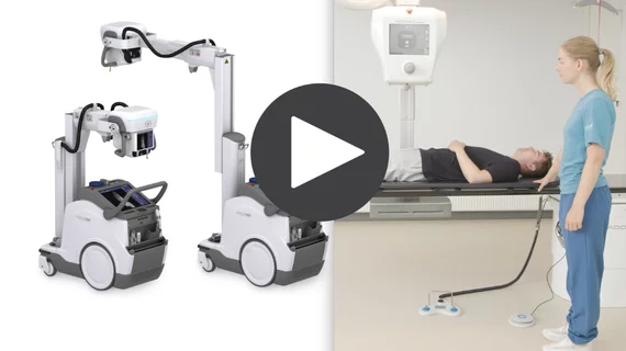
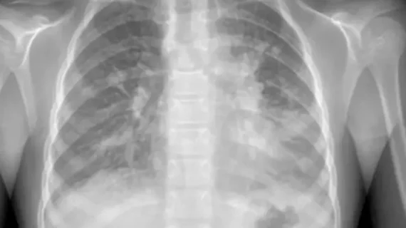
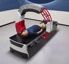
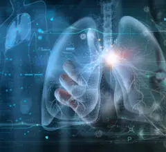
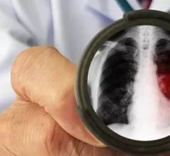
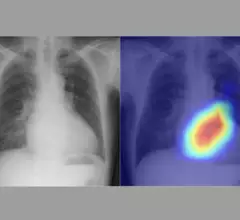
Medical X-rays have mostly converted from film, to computed radiography (CR) that used individual plates to record each X-ray digitally that then had to be uploaded into a PACS, to what is the standard-of-care today with digital radiography (DR). DR X-ray which allows immediate digital transfer of images into a PACS for immediate review. X-rays are used to diagnose fractures, bone abnormalities, lung pathologies and tumors, as well as monitor pediatric growth, plan for surgery and treat oncology patients during radiation therapy. More detailed anatomical imaging, especially soft tissue imaging, is usually sent for advanced imaging with CT or MRI. X-ray, especially mobile DR systems, are a primary use case for artificial intelligence (AI) integration. 

overnight night shift attending radiologist burnout. A new policy statement from the American College of Cardiology highlights the importance of career flexibility—including the ability to change hours or work responsibilities when necessary—for cardiologists of all ages. 

Hospital to pay False Claims Act penalty for allegedly letting unsupervised residents interpret X-rays

University of Iowa Health Care also has agreed to mandate that physicians receive training on Medicare's requirements for reimbursement in a teaching setting. 

March 23, 2023

Artificial intelligence bests ED docs, but not experienced radiologists, at detecting fractures

“This study suggests that deep learning algorithms can be useful in improving the detection of fractures," experts wrote Monday in Pediatric Radiology

March 7, 2023
Walmart Health retail care clinic

Walmart Health doubling in size, offering imaging, Epic EHR integration in 2 new states

Each of the new “state of the art” facilities will stand at about 5,750 square feet, staffed by doctors and offering the full gamut of healthcare services. 

March 3, 2023

Portable orthopedic tomosynthesis cleared for U.S. sales

The FDA has cleared U.K.-based Adaptix to market a 3D X-ray system that, according to the company, images hands, feet and elbows “at a fraction of the radiation dose and per-study cost of traditional CT.”

January 31, 2023
The Nanox ARC cold-cathode, tomosynthesis 3D X-ray system is currently pending FDA clearance. If cleared, it would be the first device of its kind to offer cold-cathode X-ray tube technology and the ability to slice through the anatomy in images similar to CT scans to aid diagnosis. The new type of tube also could greatly reduce the size and weight of X-ray systems. The vendor sees this system as key to its larger plan to address health disparities and access to imaging world-wide. #Nanox

VIDEO: Cold-cathode X-ray systems may disrupt the medical imaging market

Bhvita Jani, research manager at Signify Research, explains advances in cold-cathode X-ray tube technology and how this might represent a major shift in radiology imaging systems. 
 

January 30, 2023
Fixed digital X-ray systems have seen increased market share after a dip in 2020 when hospitals used funding to but more mobile DR systems due to COVID. The Konica-Minolta booth DR system on display at RSNA 2022. Photo by Dave Fornell

VIDEO: Trends in X-ray systems

Bhvita Jani, research manager at Signify Research, explains the technology trends she is seeing in the radiology X-ray market.

January 27, 2023
Jakob Weiss, MD, a radiologist affiliated with the Cardiovascular Imaging Research Center at Massachusetts General Hospital and the AI in Medicine program at the Brigham and Women’s Hospital in Boston, helped develop an deep learning AI algorithm that can assess a patient's biological age and risk assess patients for various diseases. #RSNA #AI #ImagingAI

VIDEO: AI predicts heart disease risk using single chest X-ray

Jakob Weiss, MD, was the lead author on a study that used AI to determine a patient's cardiovascular risks based on a standard chest X-ray.

January 12, 2023
Mahadevappa Mahesh, PhD, professor of radiology and a medical physicist at the Johns Hopkins University School of Medicine, explains a new American College of Radiology (ACR) effort to ensure that lower radiation dose X-ray images under Image Wisely and As Low as Reasonable Achievable (ALARA) meet diagnostic reading standards. He spoke to Radiology Business at the Radiological Society of North America (RSNA) 2022 meeting.

VIDEO: Making sure lower dose X-ray is still diagnostic quality

Mahadevappa Mahesh, PhD, professor of radiology and a medical physicist at the Johns Hopkins University School of Medicine, explains a new ACR effort to ensure that lower radiation dose X-ray images under Image Wisely and As Low as Reasonable Achievable (ALARA) meet diagnostic reading standards.

December 21, 2022